-
Adopt
-
Veterinary Care
Services
Client Information
- What to Expect – Angell Boston
- Client Rights and Responsibilities
- Payments / Financial Assistance
- Pharmacy
- Client Policies
- Our Doctors
- Grief Support / Counseling
- Directions and Parking
- Helpful “How-to” Pet Care
Online Payments
Emergency: Boston
Emergency: Waltham
Poison Control Hotline
-
Programs & Resources
- Careers
-
Donate Now
 By Brendan Noonan, DVM, DABVP (Avian Practice)
![]() angell.org/avianandexotic
angell.org/avianandexotic
avianandexotic@angell.org
617-989-1561
March 2023
x
x
When breaking down topics related to pet bird medicine, several unrelated yet common conditions fal l under non-infectious diseases. Many present as emergencies, given that bird illnesses can progress quickly. The focus of this article is to discuss common presentations and the management of these various conditions.
Heavy Metal Toxicity
Lead toxicity has long been recognized as the most common cause of toxicosis in birds. Lead paint is one of the most commonly encountered sources. Despite banning lead paint in 1978, many older homes still have lead paint beneath the surface. Given the capacity of flight and potential for destruction with a hook bill, many parrots become exposed without their owner’s knowledge. Stained glass windows, costume jewelry, curtain weights, grout, caulking, and linoleum are just a few other sources of toxicity noted in practice. Once ingested, the low pH of the proventriculus allows the metal to dissolve and be absorbed by the small intestine. Clinical signs encountered depend on the exposure dose.
 An acute ingestion of a large volume results in anorexia, depression, PU/PD, hematuria, paresis, and death. When a smaller amount is ingested at once or more chronic exposure over a more extended period, you are more likely to encounter weight loss, regurgitation, paresis, and nonspecific signs related to secondary immunosuppression. Typical findings on CBC include leukocytosis characterized by hetrophilia and hypochromic regenerative anemia. The chemistry panel may reveal elevations in uric acid, CK, and LDH, as well as hypoproteinemia. Radiographs can be suggestive but not definitive. As many of these patients are unstable, a “box shot” is often obtained to look for evidence of opacities more opaque than bone. It is important to note that certain non-toxic materials can appear opaque, like metal. Paint chips are not always visible and can be comparable to mineral opacity. Confirmation of the diagnosis should be performed on serum or tissue submissions. Treatment plans include basic stabilization and removal of the metal source if possible. The most commonly used chelator is calcium disodium salt of ethylenediaminetetraacetic (CaEDTA), which is administered parenterally and tends to be more effective at eliminating lead stored in bone. Stable complexes with lead are formed, which are then excreted through urine. Meso 2,3-dimercaptosuccinic acid (DMSA) is often used as the oral chelation of choice but is more effective at eliminating lead from soft tissue sources. Regurgitation has been noted even at standard therapeutic doses.
Zinc toxicity is the second most common heavy metal toxicity encountered in captive birds. Exposure is often through ingesting nuts, bolts, galvanized items, or pennies minted after 1982. Elevated zinc levels result in red blood cell hemolysis, secondary anemia, hemoglobinuria, and potentially renal failure. Nonspecific signs similar to lead toxicity, such as regurgitation, ataxia, paresis, weight loss, and death, can be encountered. Concentration levels in the pancreas and kidneys make sampling them in post-mortem exams the most useful for diagnosis. Treatment includes removing the metallic object and parenteral treatment with CaEDTA.
Inhalant Toxins
The avian respiratory system is particularly sensitive to inhalant toxins. Teflon, or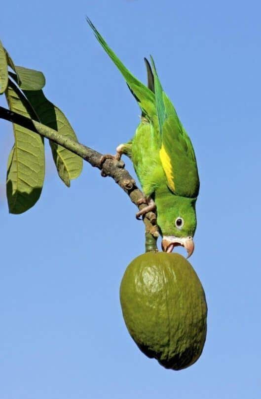 polytetrafluoroethylene (PTFE), is a common non-stick coating on pans, cooking utensils, waffle irons, drip pans, and specific tin foils. When heated above 536oF (260oC), the coating will depolymerize, releasing fumes resulting in arrhythmias or heart failure. Exposed birds will die acutely or present with severe respiratory signs and ataxia. Treatment after mild exposure has been attempted with oxygen supplementation, diuretics, bronchodilators, NSAIDs, and antibiotics. Pulmonary congestion and hemorrhage are typically noted on necropsy.
Avocado Feather Damaging Behavior
There are a few exceptions to foods suitable for parrots to share with their owners. The shortlist includes avocado, raw onion, garlic, and chocolate. Persin is the toxic substance found in an avocado tree’s leaves, fruit, bark, and seeds. The leaves have the highest concentration and tend to be the most toxic. The most common exposure is through ingestion of the fruit. Clinical signs develop between 24 to 96 hours after ingestion. Clinical signs are attributed to myocardial insufficiency and will present as dyspnea, cyanosis, cough, or acute death. Initial treatment should focus on decontamination of the crop and charcoal administration to reduce absorption. Oxygen therapy and diuretics may be useful. Post-mortem findings consist of myocardial necrosis and hemorrhage.
Feather Damaging Behavior
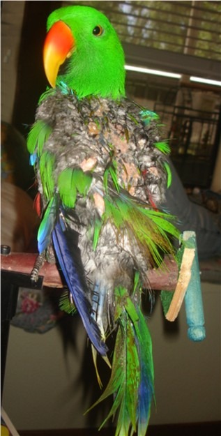 This condition encompasses a broad spectrum of behaviors that damage or remove feathers. Distinguishing changes in feather appearance and excess loss from normal molting is essential. Most parrots undergo a significant turnover of feathers one to two times per year. Light cycles, nutrition, and hormones affect the time frame. Feather damaging behavior (FDB) includes over-preening, feather chewing, and feather plucking. When damage to the skin occurs, this is referred to as self or automutilation. These abnormal behaviors are more common in birds kept in captivity. While every species has the capacity for FDB, it is more common in Cockatoos and African Greys. FDB is multifactorial but is broadly classified as either medical or behavioral. A detailed history is helpful in an acute presentation of FDB to better pinpoint contributing factors. A medical workup, including a thorough physical exam, baseline blood work, three-view radiographs, and protein electrophoresis, is suggested to get an overall picture of the patient’s health. Additional testing should be considered based on the risk factors determined in the history. Treatment is based on medical, socioenvironmental, and psychological assessment.
Egg Binding
Egg binding occurs when an egg is not laid in the expected period of time through the oviduct. Most parrots produce an egg every other day, while 80% of that time is spent in the shell gland at the distal aspect of the oviduct. This is the most common reproductive emergency and is more often seen in smaller species of parrots. Presenting complaints can include straining, weakness, dyspnea, trouble standing, and general malaise. A palpable egg is often identified within the coelomic window. Standing radiographs should be performed to confirm the diagnosis and better characterize the size, shape, and number of eggs present. A thorough history will often reveal poor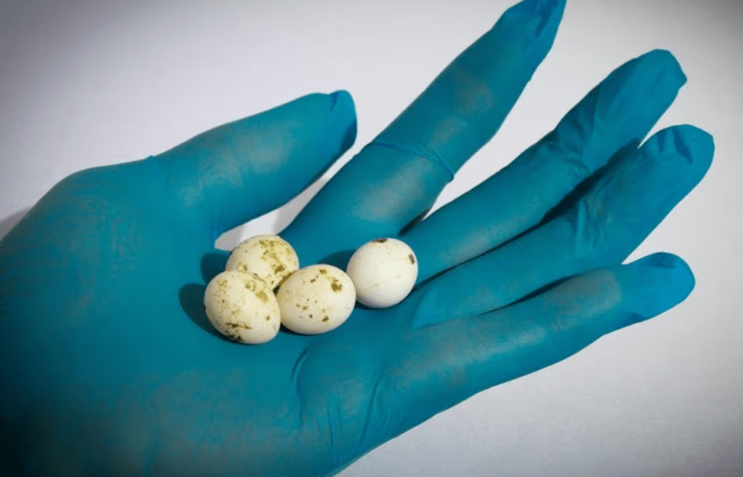 nutrition in combination with a recent history of excessive egg production. However, egg binding can occur with any prior reproductive history —implementation of supportive care, including heat support, subcutaneous fluids, calcium supplementation, and nutritional support. Pain medications or antibiotics are indicated if secondary salpingitis or tissue trauma is suspected. If no egg is produced after the first 24 to 48 hours of support, assisted delivery under general anesthesia is recommended. If the egg cannot be removed with gentle pressure, then percutaneous or transoviductal collapse can be achieved by removing the contents with a needle and syringe. Surgical intervention is recommended if the shell has become adhered or torsion is suspected.
Prolapse
Tissue protruding from the vent can be identified as cloacal, oviduct, or colon. Oviductal prolapse is often related to recent egg production, excessive straining, or adhesion of an egg to the shell gland. This tissue is best identified by longitudinal striations and a central lumen that accommodates the passage of an egg. Cloacal prolapse has been associated with behavioral and nutritional issues, infection, neoplasia, papillomatosis, and secondary reproductive complications. Evaluation for tissue damage and viability will impact prognosis. Lubrication and gentle replacement can improve viability. Lateral sutures to reduce the vent opening may be required to keep the tissue in place while the swelling resolves. This reduces the vent aperture, allowing feces to pass without tissue prolapse. Addressing the underlying etiology is necessary to prevent temporary resolution. Permanent surgical solutions should be considered for patients with recurrent prolapse secondary to behavioral or reproductive causes. Salpingohysterectomy, cloacopexy, and ventplasty should be considered. Behavioral, environmental, and nutritional modifications are necessary to improve long-term success after surgical intervention.
Iron Storage Disease
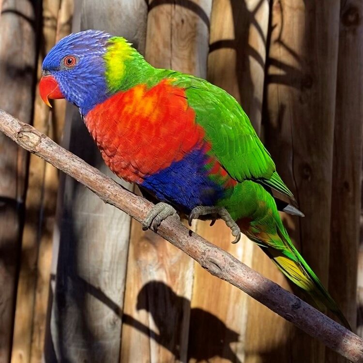 Rhamphastids, Mynahs, Birds of Paradise, and certain frugivorous parrot species (Lories, Lorikeets) are prone to hemochromatosis. Many of these species consume low-iron foods in areas with low mineral density in the soil. As a result, they are highly efficient at extracting and storing minerals. These species have no way of downregulating absorption and end up storing excessive iron in the hepatocytes as hemosiderin. Adding ascorbic and citric acid to the diet also aids translocation across the intestine and subsequent storage. Therefore, it is recommended to feed less than 100 ppm of iron per day and low-acid food items to susceptible species. This can be achieved by feeding select fruits and a commercial pellet specific for low-iron species. Clinical signs are nonspecific but generally reflect reduced hepatic function. Weight loss, ascites, dyspnea, and acute death have all been reported. Antemortem diagnosis remains difficult as blood tests are not sensitive or specific. Total iron binding capacity and serum iron levels have not been correlated with hepatic iron storage. Imaging abnormalities on survey radiographs or ultrasounds can increase suspicion but are non-confirming. Definitive diagnosis is most reliable through histology of liver biopsy using Prussian blue stains. Toxicology levels can also be determined on liver samples submitted. Treatment is based on diet modification, serial phlebotomy, and chelation. Diet modification is best used as a preventative measure, but reducing iron content to 100 ppm should be implemented. Phlebotomy is the mainstay of treatment in other species. This is contraindicated in patients who develop anemia or are diagnosed with concurrent heart disease. Chelation with deferoxamine (100mg/kg SQ Q24 x 16 weeks) has been reported and found to reduce hepatic iron storage through serial biopsies.
Atherosclerosis
Heart disease was once considered uncommon in parrot species but has since been shown to be a significant contributing factor to shorter life spans in captive species. Atherosclerosis is a chronic degenerative disease of the arteries through an accumulation of fat and cholesterol and inflammatory debris within the lumen of the vessel. Over time, this reduces the diameter and elasticity of the vessels, predisposing them to stenosis, thrombosis, and ischemia. While this is by far the most common post-mortem cardiac finding in companion psittacines, it is also suspected to be the main underlying cause for the non-infectious manifestation of heart disease. The most common places to find lesions are within the ascending aorta, brachycephalic trunks, and pulmonary arteries. Although less common, lesions such as the coronary and carotid arteries can also develop in peripheral vasculature. While any species can develop atherosclerosis, there is a predilection for African Grey parrots, Amazons, and Cockatiels. Macaws and cockatoos appear to be less susceptible. One study also found a positive correlation with advanced age, female sex, female reproductive disease, and concurrent hepatic disease. High-fat diets and reduced exercise are also contributing factors. Preventative measures are focused on diets high in pellets and fresh foods and encouraging exercise through flight and foraging activities.