-
Adopt
-
Veterinary Care
Services
Client Information
- What to Expect – Angell Boston
- Client Rights and Responsibilities
- Payments / Financial Assistance
- Pharmacy
- Client Policies
- Our Doctors
- Grief Support / Counseling
- Directions and Parking
- Helpful “How-to” Pet Care
Online Payments
Emergency: Boston
Emergency: Waltham
Poison Control Hotline
-
Programs & Resources
- Careers
-
Donate Now
 By Jennifer Peterson-Levitt, DVM, MS, DACVS
 ![]()
angell.org/surgery
surgery@angell.org
617-541-5048
April 2025
X
Overview of Primary Hyperparathyroidism
Dogs and cats have two parathyroid glands that are intimately associated with each of their thyroid glands. Generally, the cranial parathyroid gland is present on the external surface of the gland, and the more caudal parathyroid gland is located within the thyroid parenchyma. These parathyroid glands are responsible for producing parathyroid hormone (PTH), a crucial regulator of calcium levels.
Calcium homeostasis is primarily driven by PTH and calcitonin. PTH is released from Chief cells within the parathyroid glands. It causes a net increase in serum calcium by increasing calcium absorption in the kidneys and gastrointestinal tract and freeing calcium from the skeleton. PTH stimulates the kidneys to increase resorption of calcium from urine within the distal convoluted tubule and ascending loop of Henle. In the gastrointestinal tract, PTH stimulates the inactive form of vitamin D (cholecalciferol) to become active (calcitriol), resulting in increased calcium absorption in the small intestine. In addition to increasing the body’s retention of calcium from urine and ingesta, PTH also increases osteoclast activity to mobilize calcium stores within bone. In healthy animals, PTH increases serum calcium levels, and calcitonin balances the effect of PTH to prevent hypercalcemia by increasing excretion of calcium from urine, decreasing absorption from the GI tract, and inhibiting osteolysis.
In pets with Primary Hyperparathyroidism (PHPT), calcium homeostasis becomes dysregulated by excessive production of PTH from one or more parathyroid glands due to hyperplastic or neoplastic changes to the gland(s). The majority of patients are affected by parathyroid adenomas; however, parathyroid hyperplasia and carcinomas are also seen. The gold standard treatment of PHPT is surgical excision of the offending parathyroid gland(s).
Diagnosis
Primary Hyperparathyroidism should be considered as a differential diagnosis for any dog or cat presenting with clinical signs of hypercalcemia, such as polyuria, polydipsia, lethargy, muscle weakness, weight loss, decreased appetite, shaking/tremors, or urinary tract signs associated with calcium-containing urolithiasis. Interestingly, approximately one-third of patients have no clinical signs of disease, and suspicion for the disease is brought on only by detection of hypercalcemia.1 PHPT is diagnosed via blood work, with or without accompanying cervical imaging. Patients with PHPT will have a high ionized calcium level, accompanied by either a normal or elevated PTH value, and a normal parathyroid hormone-related protein (PTHrP) level. A normal PTH value is considered pathologic when seen in conjunction with hypercalcemia because an elevated calcium value should result in suppression of PTH. Patients with an elevated PTH and low ionized calcium are diagnosed with Secondary Hyperparathyroidism, which occurs as a result of a calcium-deficient diet or renal disease. Patients with a high PTHrP should be further evaluated for alternative neoplastic causes of hypercalcemia, such as Lymphoma, Mammary Carcinoma, Multiple Myeloma, and Apocrine Gland Anal Sac Adenocarcinoma (AGASACA). Although agreement between cervical ultrasound and intraoperative findings regarding the number and side of affected glands is reported to be just 65.9% and 72.3% respectively, it is valuable to confirm the presence of a nodule prior to pursuing surgery, as 3% to 6% of dogs are suspected to have accessory parathyroid tissue within the cranial thorax.2 CT scans can also be used to confirm the suspicion of one or more parathyroid nodules prior to surgery and are particularly helpful in cases where ectopic parathyroid tissue is suspected. Nuclear scintigraphy has been evaluated for use in identifying parathyroid disease in dogs, but has not proven to be useful3.
Treatment
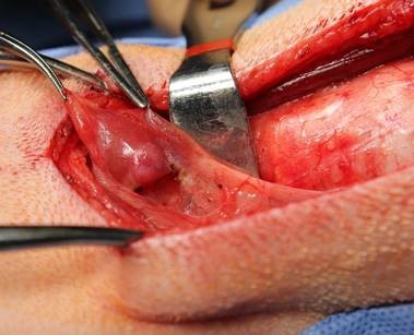
Image 1: Intraoperative photo of a Parathyroid Adenoma on the right internal parathyroid gland of a dog
Surgical excision of the offending parathyroid tissue is the mainstay of treatment for PHPT, with the majority of patients achieving resolution of hypercalcemia within 48 hours postoperatively.4 Alternative options for management of parathyroid disease, including percutaneous ultrasound-guided chemical (ethanol) and heat ablation, have been evaluated in a handful of studies; however, outcomes are less successful than those reported with surgical intervention.5 Medical management with cinacalcet may be an option for the rare dogs that are not surgical candidates or that have refractory hypercalcemia postoperatively; however, evidence is limited, and surgery remains the best option for definitive treatment.6
Following surgery, the remaining, or non-pathologic, parathyroid glands are generally atrophied as a result of chronic excessive PTH production by the parathyroid lesion(s). However, once the source of excessive PTH production is removed, the remaining parathyroid glands regain size and function as hypercalcemia resolves. In surgery, normal parathyroid glands cannot be seen or felt, and any nodular changes noted on the thyroid glands should be excised (Image 1 above).
Prophylactic calcitriol supplementation is recommended by some surgeons in an effort to avoid clinically significant hypocalcemia postoperatively; however, the literature to support this practice is mixed.4,7,8 Calcitriol has been shown to reach peak serum concentrations within three to six hours, but it takes approximately 48 hours to see an effect on calcium concentrations.4 Therefore, if clinicians plan to use calcitriol to prevent a rapid drop in calcium postoperatively, the medication should be started approximately two days prior to surgery. Calcitriol is typically initiated at a dose of 20 ng/kg/day and can then be gradually tapered over a period of two to four weeks postoperatively.4 Unlike unsupplemented patients, it is important to note that patients receiving calcitriol are unlikely to become normocalcemic within 48 hours postoperatively. These patients may experience an increase, no change, or only a mild decrease in calcium within the first 48 hours following surgery; however, they should achieve and maintain normocalcemia as the calcitriol is tapered.
While the need for prophylactic supplementation with calcitriol is questionable, patients who develop clinical signs of hypocalcemia postoperatively must be supplemented. Emergency supplementation with intravenous calcium and ECG monitoring should be performed in patients who develop seizures, muscle tremors, focal muscle twitching, and facial pawing. Following resolution of clinical signs, the patients should be transitioned to calcitriol and gradually tapered off the medication with serial monitoring of ionized calcium. Most patients ingest enough calcium from well-balanced commercial diets, but supplementation with calcium carbonate (20–50 mg/kg/day) can be considered in patients with postoperative ileus or anorexia. While supplementation is tapered, serial calcium monitoring is essential. The frequency of calcium monitoring depends on the individual patient’s calcium status, but it should be checked at least once a week. Meanwhile, owners should diligently monitor for clinical signs of hypocalcemia at home. Clinicians should aim to taper supplements at a pace that maintains serum calcium at the low end of the normal reference range, in an effort to prevent clinical signs of hypocalcemia while concurrently stimulating the function of the atrophied parathyroid glands.
Summary
PHPT is an important differential for patients with hypercalcemia. The diagnosis is confirmed by an elevated serum calcium with either a normal or elevated PTH and normal PTHrP. Surgical excision of the offending parathyroid lesion typically results in resolution of hypercalcemia within 48 hours.
References
- Feldman EC, Hoar B, Pollard R, et al. Pretreatment clinical and laboratory findings in dogs with primary hyperparathyroidism: 210 case (1987-2004). J Am Vet Med Associ. 2005;227:756-761.
- Burkhardt SJ, Sumner JP, Mann S. Ambidirectional cohort study on the agreement of ultrasonography and surgery in the identification of parathyroid pathology, and predictors of postoperative hypocalcemia in 47 dogs undergoing parathyroidectomy due to primary hyperparathyroidism. Vet Surg. 2021.50;(7):1379-1388.
- Matwichuk CL, Taylor SM, Daniel GB, et al. Double-phase parathyroid scintigraphy in dogs using technetium-99m-sestamibi. Vet Radiol Ultrasound. 2000;41(5):461-469.
- Armstrong AJ, HAuptman JG, Stanley BJ, et al. Effect of prophylactic calcitriol administration on serum ionized calcium concentrations after parathyroidectomy: 78 cases (2005-2015). J Vet Intern Med. 2018;32:99-106
- Rasor L, Pollard R, Feldman EC. Retrospective evaluation of three treatment methods for primary hyperparathyroidism in dogs. JAHA. 2007;43:70-77.
- Clark HE, Trepanier LA, Wood MW. Oral cinacalcet administration decreases serum ionize calcium and parathyroid hormone concentrations in healthy dogs. J Vet Pharmacol Ther. 2024;47(4):274-279.
- Dear JD, Kass PH, Della Maggiore AM, et al. Association of hypercalcemia before treatment with hypocalcemia after treatment in dogs with primary hyperparathyroidism. J Vet Intern Med. 2-17;31:349-35.
- Milovancev M, Schmiedt CW,. Preoperative factors associated with postoperative hypocalcemia in dogs with primary hyperparathyroidism that underwent parathyroidism: 62 cases (2004-2009). J Am Vet Med Associ. 2013;242:507-515.