-
Adopt
-
Veterinary Care
Services
Client Information
- What to Expect – Angell Boston
- Client Rights and Responsibilities
- Payments / Financial Assistance
- Pharmacy
- Client Policies
- Our Doctors
- Grief Support / Counseling
- Directions and Parking
- Helpful “How-to” Pet Care
Online Payments
Emergency: Boston
Emergency: Waltham
Poison Control Hotline
-
Programs & Resources
- Careers
-
Donate Now
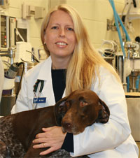 By Sue Casale, DVM, DACVS
![]()
angell.org/surgery
surgery@angell.org
617-541-5048
April 2024
Labrador Retrievers (LR) and LR crosses frequently present to veterinary surgeons for orthopedic problems, most often hind leg lameness. It is not surprising that we see a lot of LR, as they were the most popular breed of dog for 31 years before being dethroned by the French Bulldog in 2022 in the AKC rankings. Rupture of the cranial cruciate ligament (CCL) is a leading cause of hind leg lameness in dogs, accounting for about 20% of cases presenting to veterinary surgeons. (Johnson 1994) Although hip dysplasia (HD) is the most common orthopedic problem seen in dogs, many dogs (76% to 86%) with HD are asymptomatic. (Powers 2005) In a population of dogs presenting to a university hospital for hip dysplasia, 32% were found to have a rupture of the CCL as the cause of the lameness. (Powers 2005) The overall incidence of CCL rupture in dogs is about 2.5%, according to data from 2003. (Witsberger 2008) That study also showed that certain breeds are at higher risk, with Newfoundlands and Rottweilers having the highest risk. Labrador Retrievers have the third highest risk at almost 6%, about twice the average dog. Greyhounds only have a prevalence of 0.55%, making them one of the lowest-risk breeds. In that study, the prevalence of CCL rupture was calculated for each decade between 1964 and 2003, significantly increasing each decade. (Witsberger 2008) During that time, the prevalence statistically considerably increased over each period. Cruciate ligament rupture in dogs has a tremendous economic impact in the United States, with estimates that owners spent $1.32 billion on treating CCL rupture in 2003. (Wilke 2005)
ACL Disease in People
In people, anterior cruciate ligament (ACL) rupture is also a common and severe condition, with 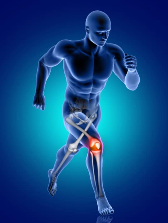 estimates of over $1 billion spent annually on surgery alone. (Flynn 2005) ACL ruptures in people have many similarities to CCL ruptures in dogs. Ruptures are most common in female athletes in their late teens or early twenties. (Sutton 2013) Greater than 70% of cases are caused by non-contact injuries and are characterized by progressive fiber tearing, which eventually progresses to complete tears. (Pfeifer 2018) People also have a high risk of contralateral rupture. ACL rupture is also a complex disease with both genetic and environmental risks. Familial risk for rupture of the anterior cruciate ligament (ACL) was first suggested in 2005. (Flynn 2005) 2.9% of patients with an ACL tear had a relative who had experienced a tear as well, which is higher than the prevalence in the overall population, making a familial predisposition likely. The same study showed that a person with an ACL tear is twice as likely to have a relative (first, second, or third-degree) who also has an ACL tear and more than twice as likely to have a first-degree relative (sibling, parent, or child) with an ACL tear. (Flynn 2005) Because the canine CCL and the human ACL are anatomically equal, the dog is often used as a model for research on cruciate ligaments in people.
CCL Anatomy
The CCL is a ligament of twisted collagenous fascicles and fiber bundles and runs cranially, medially, and distally from the femur to the tibia. (Hayashi 2004) It has a narrow mid-region, which fans out proximately and distally. Two bands make up the CCL: the craniomedial and caudolateral bands. The craniomedial band is the major contributor to the craniocaudal stability of the stifle joint. It is long and spiral but smaller than the caudolateral band. (De Rooster 2006) It is taut through range of motion, unlike the caudolateral band. The caudolateral band has a straighter course and is taut only in extension and lax in flexion. (De Rooster 2006) The craniomedial band is often torn in partial CCL tears. The blood supply to the CCL is marginal to the ligament’s core and predominantly from the surrounding soft tissue, making it vulnerable. (Hayashi 2004) There is a large amount of joint inflammation in dogs with CCL disease, which may be secondary to ligament rupture; however, in 25% of cases, synovitis is present prior to ligament fraying. (Bleedhorn 2011) This may indicate that synovitis may cause ligament degeneration in some dogs. (Bleedhorn 2011, Muir 2011) CCL rupture in dogs is a syndrome of progressive fiber tearing in the presence of knee synovitis. Joint inflammation, mechanical overloading, ligament micro-injury, and ischemia all result in diminished typical crimped structure of collagen fibrils and disruption of ligament fascicles. (Hayashi 2004, De Rooster 2006) The increased collagen remodeling leads to increased laxity and progressive degenerative joint disease. Most cases involve a mid-substance rupture, which is the narrowest region of the ligament and has the weakest blood supply. (Bennett 1988, Hayashi 2004)
Risk Factors for CCL Disease
CCL tears typically occur as a non-contact injury during normal activity. (Pfeifer 2018) Normal running,  fetching, and playing with other dogs should not be enough force to rupture a healthy ligament. (Buote 2006) High-level activities such as agility, dock jumping, racing, and lure coursing do not increase the risk of CCL rupture. (Sellon 2022) In fact, competing frequently and participating in more challenging events was associated with a lower risk of CCL rupture, while novice-level agility was associated with an increased risk. Other studies also found that activity level was not a risk for CCL rupture in Labrador Retrievers. (Terhaar 2020) That same study and a second study showed that dogs who were obese or overweight, on a weight management plan, or fed premium dog food were at increased risk. (Lampman, 2003) CCL rupture is a multifactorial disease; many studies have evaluated risk factors. Age, sex, neuter status, weight, conformation, and breed have all been implicated as influencing the risk; however, many studies have found contradictory results. Early studies on the CCL found microscopic degenerative disease in the cranial and caudal cruciate ligament as dogs age. (Zahm 1965, Vassuer 1986) The degeneration progressed, and no attempt was made at repair. (Vassuer 1986) This degeneration leads to the weakening of the ligaments in older dogs but does not explain the ruptures seen in young dogs. In 1988, it was suggested that there is a syndrome of CCL disease in younger large breed dogs where dogs less than four years of age and greater than 22kg experience partial ruptures that progress to complete tears over time. (Bennett 1988)
CCL Disease and Genetics
A decade later, researchers started investigating the breeds experiencing early CCL tears. Newfoundlands were found to have an increased risk for CCL, while Golden Retrievers and German Shepherds had decreased risk. (Duval 1999) This suggested heritability in cruciate disease. Greyhounds rarely rupture their CCL, so they have served as controls in many studies. Both Rottweilers and LR were found to have weaker CCLs when compared to greyhounds. (Wingfield 2000, Comerford 2005) Numerous studies describe a high risk of bilateral tears in young, high-risk breeds. Increased weight bearing on the contralateral side is not the cause of bilateral tears, as dogs with experimentally transected cruciate ligaments had normal contralateral joints after being followed for over two years.(Campbell1982) Bilateral rupture on initial presentation has been reported in as many as 27% of dogs. Dogs presenting with unilateral disease have a 37% to 48% risk of tearing the other side. (Doverspike 1993, Buote 2009, Cabrera 2009) Degenerative change or joint effusion in the contralateral stifle is associated with an even higher risk (59% to 61%), especially in younger dogs. (Fuller 2014) Dogs over eight years of age are less likely to experience a contralateral tear (19%). (Murphy 2023) Labrador Retrievers had an even lower risk after eight years of age with only 6% rupturing the contralateral side. (Cook 2020) A genetic basis for CCL rupture was first suggested in 2003 when pedigree analysis showed genetic heritability in both boxers and Newfoundlands. (Nielen 2003, Wilke 2006) In 2009, chromosomal regions associated with CCL rupture were identified, and in 2014, three chromosomal regions associated with CCL disease were found in Newfoundlands, confirming a genetic basis in that breed. (Baird 2014) That same year, single nucleotide polymorphisms (SNPs) were examined in four breeds: Newfoundlands, Rottweilers, LR, and Staffordshire Terriers. (Baird 2014) SNPs are a substitution of a single nucleotide at a specific position in the genome that is present in varying numbers within a population. They may influence the development of a disease and control how a patient will respond to pathogens or medications. The authors looked at 196 SNPs across 28 genes and found 17 were associated with the CCL, most within collagen genes. Genes involved in ligament strength, stability, and extracellular matrix formation were all associated with rupture of the CCL. (Baird 2014) The authors concluded that the structure and strength of the CCL may be compromised by these mutations, leading to an increased risk of CCL rupture. (Baird 2014) SNP-based heritability in LR was investigated, and heritability ranged from 0.55 to 0.886. (Cook 2020) This means that 55% to 89% of the risk of developing CCL rupture is genetic in LR, with environmental factors accounting for the remainder. That study also found that less than 6% of LR ruptured their CCL after eight years of age, so dogs older than eight were used as controls. (Cook 2020) Another study found that yellow LR had an increased risk of CCL rupture while black LR had a decreased risk. (Terhaar 2020) This was the first time coat color influencing the risk of CCL was described. That same group analyzed 679 SNPs and found that multiple SNPs are associated with both CCL disease and coat color. (Lee 2023) Inheritance of coat color is controlled by two genes, MC1R and TYRP1. A mutation within MC1R is responsible for the yellow color in LR. Dogs that are homozygous for the MC1R mutation produce pheomelanin, which creates a yellow coat color. In contrast, dogs with at least one gene without the mutation will produce eumelanin, resulting in a black or chocolate coloring. (Lee 2023) It is possible that the selection for coat color inadvertently selected risk variants for other phenotypes, including CCL disease. Coat color does influence behavior and other disease processes, such as skin and ear disease, which is statistically more frequent in chocolate LR. ( McGreevy 2018). Chocolate color is also associated with a higher body condition score. (Wallis 2023)
Testing for CCL Disease
Recently, genetic testing became available specifically for LR and CCL disease. The test uses SNP markers 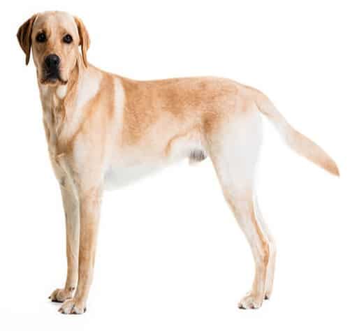 and sex to determine risk so it can be performed at any age. The test is currently only available for LR, but additional breeds should eventually be available. Many genetic markers across the genome are examined. Although each variant may have a small to moderate effect, they act additively, and a higher number of DNA risk variants increases the genetic risk of developing CCL rupture. The results are reported as “predicted to be a case” if marker genotypes associated with CCL disease are present and “predicted to be control” if marker genotypes protective from CCL rupture are present. Patients predicted to be a case are very likely to rupture their CCL, while dogs “predicted to be control” are unlikely to rupture their CCL. The test is available from the University of Wisconsin Comparative Genetics and Orthopedic Research Lab. Blood or saliva can be submitted, and results take approximately four to six weeks. The test costs about $250 and is 98% accurate. It can be ordered through genetics@vetmed.wisc.edu. The most significant benefit of this test is for breeders, and it should be completed along with screening for eyes, elbows, and hips; only low-risk dogs should be bred. Puppies intended for hunting or athletic work should also be screened prior to investing in training. Pets may also benefit from testing so lifestyle changes can be undertaken and treatment can be pursued with early signs of disease prior to the development of osteoarthritis. The impact of a positive test on insurance coverage may need to be considered.
Conclusion
CCL rupture is a disease that is the leading cause of hind limb lameness in dogs, with numbers steadily increasing in frequency. In Labrador Retrievers, most of the risk of CCL rupture is genetic, and testing breeding dogs will help select dogs with lower risk. Most CCL ruptures occur in dogs under eight years of age, and LR have a decreased risk after eight years of age compared to other dogs.
References
- Baird AEG, Carter SD, Innes JF, et al. Genetic basis of cranial cruciate ligament rupture (CCLR) in dogs. Connect Tissue Res 2014;55(4):275-281.
- Baird AEG, Carter SD, Innes JF, et al. Genome-wide association study identifies genomic regions of association for cruciate ligament rupture in Newfoundland dogs. Animal Genetics 2014;45:542-549.
- Bennett D, Tennant B, Lewis DG, et al. A reappraisal of anterior cruciate ligament disease in the dog. J Small Anim Pract 1988: 29:275-297.
- Binversie EE, BakerLA, Engelman CD, et al. Analysis of copy of number variations in dogs implicates genomic structural variation in the development of anterior cruciate ligament rupture. PLOS one, December 31, 2020 doi.org/10.1371/journal.pone.0244075.
- Bleedhorn JA, Greuel EN, Manley PA et al. Synovitis in dogs with stable stifle joints and incipient cranial cruciate ligament rupture: a cross-sectional study. Vet Surg 2011;40:531-543.
- Buote N, Fusco J, Radasch R. Age, tibial plateau angle, sex and weight as risk factors for contralateral rupture of the cranial cruciate ligament in Labradors. Vet Surg 2009; 38:481-489.
- Cabrera SY, Owen TJ, Mueller MG, Kass PH. Comparison of tibial plateau angles in dogs with unilateral versus bilateral cranial cruciate ligament rupture: 150 cases (2000-2006). JAVMA 2008;232(6):889-892.
- Campbell JR, Duff SRI, Gilberson EMM. The effect on the contralateral stifle joint of sectioning of the cranial cruciate ligament in the dog. J Small Anim Pract 1982;23:511-516.
- Comerford EJ, Tarlton JF, Innes JF, et al. Metabolism and composition of the canine anterior cruciate ligament relate to differences in knee joint mechanics and predisposition to ligament rupture. J Ortho Res 2005;23:61-66.
- Cook SR, Conzemius MG, McCue ME, Ekensted KJ. SNP-based heritability and genetic architecture of cranial cruciate ligament rupture in Labrador Retrievers. Anim Gen 2020; 51:824-828.
- Cunningham DP, Mostafa AA, Gordan-Evans WJ, et al. Factors contributing to the variability of a predictive score for cranial cruciate ligament deficiency in Labrador Retrievers. BMC Vet Research 2017;13:235.
- DeRooster H, DeBruin T, VanBree,H. Morphologic and functional features of the canine cranial cruciate ligaments. Vet Surg 2006;35:769-780.
- Duval JM, Budsberg SC, Flo GL, et al. Breed, sex and body weight as risk factors for rupture of the cranial cruciate ligament in young dogs. JAVMA 1999;215:811-814
- Fitch RB, Beale BS. Osteochondrosis of the canine tibiotarsal joint. Vet Clin North Am 1998;28(1):95-113.
- Fuller MC, Hayashi K, Bruecker KA, et al. Evaluation of the radiographic infrapatellar fat pad sign of the contralateral stifle joint as a risk factor for subsequent contralateral cranial cruciate ligament rupture in dogs with unilateral rupture: 96 cases (2006-2007). J Am Vet Med Assoc 2014; 244:328-338.
- Flynn RK, Pedersen CL, Birmingham TB, et al. The Familial predisposition toward tearing of the anterior cruciate ligament. A case control study. Am J Sports Med 2005;33(1):23-28.
- Gibbons SE, Macias C, Tonzing MA, et al. Patellar luxation in 70 large breed dogs J Small Anim Pract 2006;47:3–9
- Grierson J, Asher, L, Grainger K. An investigation into risk factors for bilateral cranial cruciate ligament rupture. Vet Comp Orthop Traumatol 2011;24:192-196.
- Guthrie JW, Keeley BJ, Maddock SR, et al. Effect of signalment on the presentation of canine patients suffering from cranial cruciate ligament disease. J Sm Anim Pract 2012;53:273-277.
- Hayashi K, Manley PA, Muir P. Cranial cruciate ligament pathophysiology in dogs with cruciate disease: a review. J Am Anim Hosp Assoc 2004;40:385-390.
- Hayashi K, Frank JD, Dubinsky C, et al. Histologic changes in ruptured canine cranial cruciate ligament. Vet Surg 2003;32:269-277.
- Hayes AG, Boudrieau RJ, Hungerford LL. Frequency and distribution of medial and lateral patellar luxation in dogs: 124 cases (1982-1992). JAVMA 1994;205(5):716-720.
- Johnson JA, Austin C, Breur GJ. Incidence of canine appendicular musculoskeletal disorders in 16 veterinary teaching hospitals form 1980 through 1989. Vet Comp Orthop Traumatol 1994;7:56-69.
- Lampman TJ, Lund EM, Lipowitz AJ. Cranial cruciate disease: current status of diagnosis, surgery, and risk for disease. Vet Comp Orthop Traumatol 2003;16:122-126.
- Lee BT, Baker LA, Momen M, et al. Identification of genetic varients associated with anterior cruciate ligament rupture and AKC standard coat color in the Labrador Retriever. BMC Vet Research 2023;24:60.
- McGreevy PD, Wilson BJ, Mansfield CS, et al. Labrador retrievers under primary veterinary care in the UK: demography, mortality and disorders. Canine Gen Epidem 2018;5:8.
- Muir P, Schwartz Z, Malek S, et al. Contralateral cruciate survival in dogs with unilateral non-contact cranial cruciate ligament rupture. PLOS One 2011;6(10):e25331.
- Murphy CL, Niles J, Radasch, RM. The prevalence and risk factors of contralateral cranial cruciate ligament rupture in medium-to-large (>15kg) breed dogs 8 years of age or older Vet Comp Orthop Traumatol 2024;37:8-12.
- Pfeifer CE, Beattie PF, Sacko RS, hand A. Risk factors associated with non-contact anterior cruciate ligament injury: a systemic review. Int J Sports Phys Therap 2018;13(4):575-587.
- Powers MY, Martinez SA, Lincoln JD, Temple CJ. Prevalence of cranial cruciate ligament rupture in a population of dogs with lameness previously attributed to hip dysplasia: 369 cases (1994-2003). JAVMA 2005;227(7):1109-1111.
- Sellon DC, Marcellin-Little DJ. Risk factors for cranial cruciate ligament rupture in dogs participating in canine agility. BMC Vet Research 2022;18:39
- Smith KD, Hayashi K, Clements DN, et al. Variation in the quantity of elastic fibres with degeneration in cranial cruciate ligaments form Labrador Retrievers. Vet Comp Orthop Traumatol 2017;30:398-402.
- Sutton KM, Bullock JM. Anterior cruciate ligament rupture: differences between males and females. J Am Acad Orthop Surg 2013;21(1):41-50
- Terhaar HM, Muir P, Baker LA, et al. Contribution of habitual activity to cruciate ligament rupture in Labrador Retrievers. Vet Comp Orthop Traumatol 2020;33(2):82-88.
- Vassuer PB, Pool RR, Arnoczky RR, Lau RE. A correlative biochemical and histological study of the cranial cruciate ligament in dogs. 1986;Am J Vet Res;46:1842-1854.
- Wallis NJ, Sumanasekera NT, Raffan E. Obesity risk factors in British Labrador retrievers: Effect of sex, neuter status, age, chocolate coat colour and food modivation. Vet Rec 2023;e3410.
- Wilke VL, Robinson DA, Evans RB, et al. Estimate of the annual economic impact of treatment of cranial cruciate ligament injury in dogs in the United States. JAVMA 2005;227(10):1604-1607.
- Wilke VL, Zhang S, Evans RB, et al. Identification of chromosomal regions associated with cranial cruciate ligament rupture in a population of Newfoundlands. AJVR 2009;70(8):1013-1017.
- Wilke VL, Conzemius MG, Koinghorn BP, et al. Inheritance of rupture of the cranial cruciate ligament in Newfoundlands. JAVMA 2006;228(1):61-64.
- Wingfield C, Amis AA, Stead AC, Law, HT. Cranial cruciate stability in the Rottweiler and racing greyhound: an in vitro study. J Small Anim Pract. 2000;41:193-197.
- Wingfield C, Amis AA, Stead AC, Law HT. Comparison of the biomechanical properties of Rottweiler and racing greyhound cranial cruciate ligaments. J Sm Anim Pract 2000;41:303-307.
- Witsberger TH, Villamil JA, Schultz LG, et al. Prevalence of and risk factors for hip dysplasia and cranial cruciate ligament deficiency in dogs. JAVMA 2008;232(12):1818-1824.
- Zahm H. Die ligament decessata in gesunden und arthrotischen kniegelenk des hundes. Kleintierpraxia 1965;10:28-47.
- Zeltzman PA, Pare B, Johnson GM, et al. Relationship between age and tibial plateau angle in dogs with cranial cruciate rupture. J Am Anim Hosp Assoc 2005;41:117-120.