-
Adopt
-
Veterinary Care
Services
Client Information
- What to Expect – Angell Boston
- Client Rights and Responsibilities
- Payments / Financial Assistance
- Pharmacy
- Client Policies
- Our Doctors
- Grief Support / Counseling
- Directions and Parking
- Helpful “How-to” Pet Care
Online Payments
Emergency: Boston
Emergency: Waltham
Poison Control Hotline
-
Programs & Resources
- Careers
-
Donate Now
angell.org/internalmedicine
(617) 522-7282
internalmedicine@angell.org
Tracheal Stenting
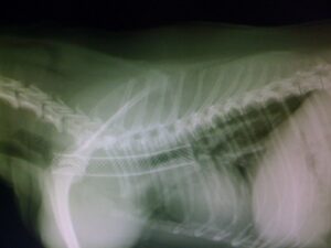
Tracheal collapse is a common, progressive, degenerative disease of the cartilage rings that primarily affects small- and toy-breed dogs. This condition is the result of low cellularity and decreased glycosaminoglycan and calcium content of the cartilage. These alterations lead to dynamic airway collapse during respiration. Luminal collapse of the tracheal dorsal membrane, mainstem bronchus collapse, and other lower airway conditions can also be seen in conjunction with tracheal collapse, further contributing to clinical signs. The collapse can be focal or diffuse throughout the length of the trachea.
Affected animals present with signs ranging from mild, intermittent “honking” to severe respiratory distress. An initial diagnosis is often made based on breed and the characteristic cough. Testing to document tracheal collapse includes thoracic films, tracheoscopy, or fluoroscopy. Identification of the extent of the trachea affected (extra-thoracic, thoracic inlet, intra-thoracic, or diffuse collapse) as well as whether mainstem bronchus collapse is present, help in determining a treatment plan and prognosis.
Initial treatment is often aimed at controlling the cough with medications and by maintaining a good body weight, using a harness, limiting episodes where there may be excitement, and decreasing environmental allergens. Medications used include anti-tussives, steroids (anti-inflammatory doses), bronchodilators (concurrent bronchial disease), antibiotics if there is a secondary infection, and antihistamines. Many patients will do very well with medical management; however, a percentage will continue to have clinical signs, sometimes severe. Tracheal stenting is an option in dogs that have failed medical management. Candidates for stenting are often those that are older, those whose pathology involves the intrathoracic or the entire trachea, those that are extremely small (in which prosthetic rings may be more complex), those that present intubated in crisis or are unable to be extubated due to severe respiratory distress, and those whose owners prefer a non-surgical approach to therapy. It is important to emphasize to clients that tracheal stenting is an adjunct treatment to medical management and not a replacement therapy.
Tracheal stenting is a minimally invasive procedure used to re-establish patency, using a self-expanding metallic stent (nitinol) which holds the trachea open through outward radial force. Stents are most often placed for tracheal collapse, but may also be placed when tracheal tumors are narrowing the lumen. Unlike tracheal rings, stents can be used for tracheal collapse at any anatomic level (cervical, thoracic inlet, intra-thoracic). Measurements and stent placement are performed under general anesthesia with fluoroscopic guidance and are relatively quick. About 75–90% of animals improve with stent placement. If there is mainstem bronchus collapse, the response rate is lower (50–75%). Complications during the procedure include subcutaneous emphysema and incorrect placement location. These complications can be life-threatening, but are considered uncommon. Provided there are no immediate complications, most patients are able to be discharged the following day. Cough control is extremely important in the first few weeks following stent placement, but medications are tapered over time to lowest effective dosing. Long-term complications include stent shortening over time, excessive inflammatory tissue formation around the stent, stent fracture, and progressive disease. Excessive granulation tissue often responds to treatment with steroids, and stent fractures can be corrected by the placement of another stent within the fractured stent.
Urethral Stenting
Urethral stenting is also currently available at Angell. Palliative urethral stenting offers relief to dogs with urinary obstruction due to infiltrative disease associated with the bladder, prostate, and other intra-pelvic structures. The ”gold standard” therapies for urethral neoplasia should still be discussed with owners, including systemic chemotherapy, as uretheral stenting only addresses clinical signs. Also, the stents used commonly are self-expanding nitinol. A contrast urethrogram is performed prior to stent placement to obtain measurements, and deployment follows, using fluoroscopic imaging to ensure accurate positioning. Urinary incontinence is the biggest complication. Incontinence rates, of any degree, in a recent study ranged from about 45% (females) to 75% (males), with about 25% of dogs being severely affected. Owners should be counseled on this complication and the palliative nature of the stent prior to placement.
For more information or referral for a tracheal or urethral stent, please contact Dr. Shawn Kearns or Dr. Maureen Carroll at (617) 522-7282 or internalmedicine@angell.org.


