-
Adopt
-
Veterinary Care
Services
Client Information
- What to Expect – Angell Boston
- Client Rights and Responsibilities
- Payments / Financial Assistance
- Pharmacy
- Client Policies
- Our Doctors
- Grief Support / Counseling
- Directions and Parking
- Helpful “How-to” Pet Care
Online Payments
Referrals
- Referral Forms/Contact
- Direct Connect
- Referring Veterinarian Portal
- Clinical Articles
- Partners in Care Newsletter
CE, Internships & Alumni Info
CE Seminar Schedule
Emergency: Boston
Emergency: Waltham
Poison Control Hotline
-
Programs & Resources
- Careers
-
Donate Now
 By Klaus Loft, DVM
www.angell.org/dermatology
dermatology@angell.org
617 524-5733
Sebaceous adenitis (SA) is a relatively uncommon, immune-mediated dermatosis causing a crusting cornification abnormality, which in turn can lead to progressing alopecia with tightly adherent scales and crusts (see Figure 1).

Figure 1
The etiology is somewhat unclear, but the inflammatory focus is the sebaceous glands and ducts, which affect the hair follicle’s ability to regrow hair. SA often presents with a significant change in the pet’s exterior and coat texture and quality. The hair coat in affected areas often changes both in consistency and color after being damaged in the process (e.g., apricot to dark brown, or soft to wired hair) (see Figure 2). The first reports in the veterinary literature by Scott et al. and Rosser et al. plus White et al.1, 2, 3 all reported that moderate to severe crusting dermatosis in Standard Poodles and Akitas and the histopathological samples from these dogs were devoid of sebaceous glands. SA is generally considered to be a dermatological condition not existing clinically prior to the early 1980s, and reviews of banked histopathological tissues have not revealed any evidence of SA.4, 5
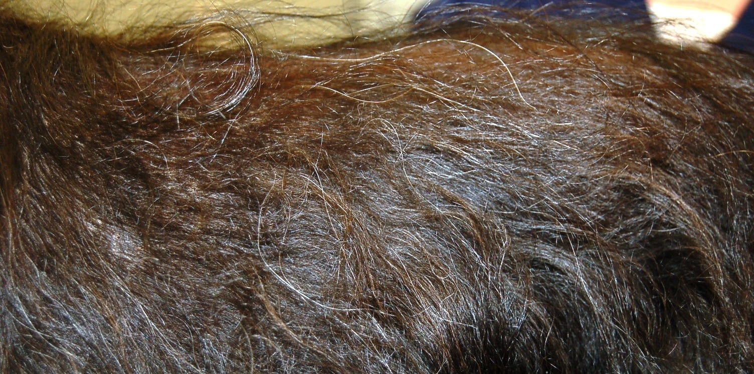
Figure 2
Affected dogs present with a mildly to moderate pruritic crusting skin condition and suffer from intermittent bacterial or yeast-related pyoderma (see Figure 3). Certain lines of Standard Poodles have appeared more heavily affected, and a voluntary screening program implemented within the breed has resulted in the condition being more or less eradicated in this breed, but over the last 10+ years the condition has been seen in a longer list of breeds, including the Husky, Bichon Havanese6 and Vizla.7,8 An autosomal recessive mode of inheritance with a variable expression has been proposed in the Standard Poodle9, 10, 11 and an autosomal recessive mode of inheritance is suggested as the reason for the Akita7 being more frequently affected. The condition has been diagnosed by the Angell Dermatology Service in the following breeds: Springer Spaniel, Welsh Corgi, German Shepherd dog, Labrador Retriever, Bernese Mountain dog, Cockapoo, Goldendoodle, and Labradoodle. A similar but even rarer condition has been reported in humans, cats, horses, and rabbits.3, 4, 5, 6
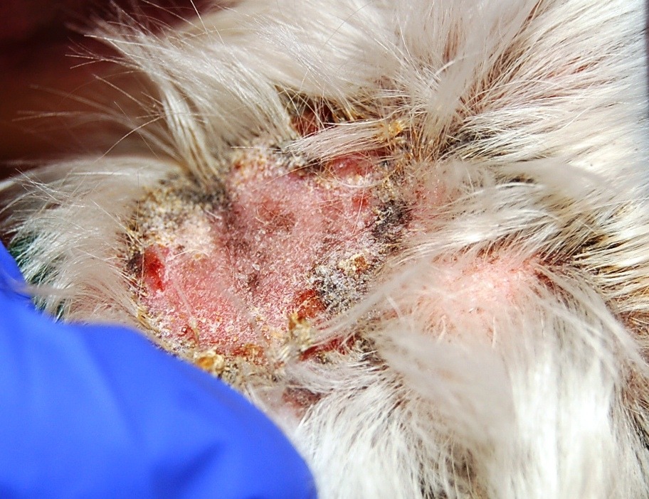
Figure 3
SA is a largely cosmetic condition with minimal or no systemic symptoms. It generally does not cause pruritus or pain, but pets with this condition are prone to secondary bacterial and/or yeast infections that can cause pruritus, further inflammation, and potential additional hair follicle destruction. Secondary infections should be treated to avoid any further damage to the hair follicles. In general, dogs affected by this condition should not be used for breeding, as there appears to be a potential genetic predisposition.
Numerous treatments and supportive therapies are used in the medical management of the primary disease and treatment for secondary infections. Treatment for SA will typically include one or more of the following oral therapies:9, 10, 11, 12, 13, 14
VITAMIN A: This vitamin is known to have a potential teratogenic effect in high dosages, and does have the ability to slow down the cell turnover in the keratinization process. Therefore, Vitamin A can assist by reducing the severity of the intense scaling. The use of this vitamin supplement can occasionally cause a dry eye condition, keratoconjunctivitis sicca (KCS), so it is important to have a tear production test done prior to starting this treatment.
PRIMROSE OIL AND ESSENTIAL FATTY ACIDS (EFA): Supplementing the diet can assist in the capacity of anti-inflammatory precursors to rebuild the skin and can have an antioxidant-like effect within the affected tissue. A diet rich in omega-3 and -6 fatty acids, such as Hill’s Science Diet Sensitive Skin and Royal Canine Skin Support, can be very helpful in making skin more resilient to trauma and skin infections. Alternatively, fatty acids supplements such as 3V Caps, AllerDerm, or Derm Caps can be added to any balanced diet.
SYNTHETIC RETINOIDS (tretinoin and isotretinoin): Several authors have used this treatment modality for SA to reduce the crusting and scaling following the adnexal destruction of the sebaceous glands. The number of cases in the literature is relatively low, and the concerns for KCS and teratogenicity are significantly higher than in Vitamin A. Therefore, synthetic retinoids are not a first-line option in most cases.
CYCLOSPORINE (ATOPICA): This medication helps modulate the immune response against the targeted tissue in SA. The cost of long-term or lifelong dosing is often a significant concern. The most common possible side effects of this medication are vomiting and diarrhea and are usually self-limiting, but can be so significant that treatment will have to be stopped. We recommend giving this medication on an empty stomach for more even absorption, but if gastrointestinal side effects do occur, it can be given with food.
There are also topical therapies that may be prescribed, such as:
ALPHA KERI BATH OIL (mineral oil mix): This occlusive treatment will make it possible to separate the very adherent sebaceous material from the coat and reduce the odor and risk for secondary pyoderma. This treatment is often time-consuming and fairly messy!
PROPYLENE GLYCOL 70%: This topical product is usually prepared by a human pharmacy and can be applied with a spray bottle to help moisturize the skin every 24–72 hours for 2–3 weeks. After this initial period, application can be reduced to the lowest possible interval, but at least weekly use is needed.
This disease can be a frustrating condition for clinicians, pet owners, and pets, especially as we see an increase in the popularity of “designer breeds” with the Poodles in the mix, and the spontaneous occurrence of the condition in more types of dog breeds. In the future, gene mapping might assist in finding a specific genetic defect and the availability of more specific therapies.
For more information about canine sebaceous adenitis or Angell’s Dermatology Service, please call 617 524-5733 or e-mail dermatology@angell.org. You can also reach Dr. Loft at keloft@angell.org.
References
1 Scott DW, Miller WH Jr, Griffin CE: Muller and Kirk’s Small Animal Dermatology, 6th ed. Philadelphia: W.B. Saunders Co., 2001: p. 1140–1146.
2 Rosser E, Dunstan R, Breen P et al.: “Sebaceous adenitis with hyperkeratosis in the standard poodle: a discussion of 10 cases.” JAAHA, 1987; 23: p. 341.
3 White S, Linder K, Schultheiss P et al.: “Sebaceous adenitis in four domestic rabbits (Oryctatagus cuniculus).” Veterinary Dermatology 2000; 11: p. 53–60.
4 Gross T, Ihrke P, Walder E, Affolter V: “Sebaceous adenitis.” In: Skin diseases of the dog and cat, Clinical Histopathological Diagnosis. 2nd ed. Oxford: Blackwell Publishing, 2005, p. 186–88.
5 Yager J, Willcock B, eds. Surgical pathology of the dog and cat. London: Mosby Year Book Inc., 1994; p. 197–198.
6 Osborne C: “Sebaceous adenitis in a 7-year-old Arabian gelding.” Canadian Veterinary Journal 2006; 47: p. 583–6.
7 Zur G, Botero-Anug A: “Severe Ulcerative and Granulomatous Pinnal Lesions with Granulomatous Sebaceous Adenitis in Unrelated Vizslas.” JAAHA, 47:6 Nov./Dec. 2011, p. 455–60.
8 Hernblad Tevell E, Bergvall K, Egenvall A: “Sebaceous adenitis in Swedish dogs, a retrospective study of 104 cases.” Acta Veterinaria Scandinavica 2008, 50:11, p. 1–8.
9 Frazer M, Schick A, Lewis T, Jazic E: “Sebaceous adenitis in Havanese dogs: a retrospective study of the clinical presentation and incidence.” Veterinary Dermatology, 2010, 22, p. 267–274.
10 Rosser E: “Therapy for sebaceous adenitis.” In: Bonagura JD, ed. Kirk’s Current Veterinary Therapy XIII. Philadelphia: W.B. Saunders, 2000: p. 572–3.
11 Rosser E: “Sebaceous adenitis.” In: Kirk RW, Bonagura JD, eds. Current Veterinary Therapy XI: Small Animal Practice. Philadelphia: W.B. Saunders, 1992: p. 534–6.
12 Rosser E: “Sebaceous adenitis.” In: Kirk RW, Bonagura JD, eds. Current Veterinary Therapy XIV: Small Animal Practice. Philadelphia: W.B. Saunders, 2009, p. 449–453.
13 Sousa C: “Sebaceous adenitis.” Veterinary Clinics of North America: Small Animal Practice 2006; 36: p. 243–9.
14 Linek M, Boss C, Haemmerling R, Hewicker-Trautwein M, Mecklenburg L: “Effects of cyclosporine A on clinical and histologic abnormalities in dogs with sebaceous adenitis.” JAVMA, Vol. 226, No. 1, January 1, 2005, p. 59–64.