-
Adopt
-
Veterinary Care
Services
Client Information
- What to Expect – Angell Boston
- Client Rights and Responsibilities
- Payments / Financial Assistance
- Pharmacy
- Client Policies
- Our Doctors
- Grief Support / Counseling
- Directions and Parking
- Helpful “How-to” Pet Care
Online Payments
Referrals
- Referral Forms/Contact
- Direct Connect
- Referring Veterinarian Portal
- Clinical Articles
- Partners in Care Newsletter
CE, Internships & Alumni Info
CE Seminar Schedule
Emergency: Boston
Emergency: Waltham
Poison Control Hotline
-
Programs & Resources
- Careers
-
Donate Now
 By Brendan Noonan, DVM, DABVP
![]()
angell.org/avianandexotic
avianandexotic@angell.org
617-989-1561
January 2022
Through our active emergency service, the most common presentation for rabbits is a change in appetite. Most owners know that rabbits should be steadily eating and defecating throughout the day. When these habits change, either acutely or over several days, evaluation by a veterinarian is warranted. Liver lobe torsion (LLT) has become increasingly recognized as a differential in rabbits presenting non-specific gastrointestinal stasis signs. Early identification of an underlying cause is crucial to developing a treatment plan and prognosis.
Gastrointestinal stasis acts as an umbrella term for decreased appetite or fecal production. This syndrome can also be described as ileus since gastrointestinal contractions have slowed or diminished. Owners will report that fecal pellets are smaller, dryer, or fewer in number over several days. Appetite may also be diminished, and some patients will only accept treats or their favorite food items. A variety of underlying causes can contribute to the symptoms displayed, and these different etiologies will alter the implemented treatment plan. Diet, underlying health conditions, pain, or stress can all contribute to the onset of gastrointestinal stasis. One day of anorexia and decreased fecal production is the most common complaint about rabbits later identified with liver lobe torsion.
With the high emergency caseload at Angell, we use temperature as one of the main determining factors for whether hospitalization or outpatient therapy is recommended. When the emergency service triages a rabbit for decreased appetite, body temperature is one of the most important early signs. All hypothermic rabbits are provided with immediate heat support using heated blankets, heated discs, incubators, or forced air blankets. Patients presenting with gastrointestinal stasis have a normal core temperature (100.5-103.5) or mild hypothermia (98-100°F). Rabbits suffering from GI obstruction may be in hypovolemic or decompensatory shock and are often profoundly hypothermic (<98°F). In our experience, most rabbits later identified as liver lobe torsions will present with normal temperatures. Given the potential to recommend outpatient therapy for a rabbit with a 24-hour history of decreased appetite, normal temperature, and mild abdominal discomfort during palpation, blood work is routinely offered. In some instances, blood work has identified the suspicion of LLT in patients who have left the hospital and need to come back for further workup and support.
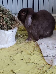
In a groundbreaking retrospective study, Graham et al. followed 16 cases over five years at Angell (2007-2012), highlighting the workup and outcome of confirmed LLTs. While this condition is still uncommon, their work has recognized a potentially overlooked diagnosis with a non-specific presentation. Angell has identified and treated at least 15 cases in 2021 alone and, at one point over the summer, had eight cases in a six-week time frame. The average age of rabbits presenting is five with no clear sex predilection. Graham et al. identified Mini Lops as the most common breed comprising 11 of the 16 cases reported. While lop rabbits continue to be over-represented in our caseload, we have seen this condition affect rabbits of numerous other breeds and sizes. LLT has been identified in countless other species, with dogs most common in veterinary medicine. The etiology of LLT is still unknown, but predisposing factors include trauma, congenital absence of hepatic ligaments, organ enlargement, or other hepatic pathology. The caudate liver lobe is the most commonly affected in rabbits, theoretically due to a long tapered attachment to the hilar region of the liver. Graham et al. did identify cases where the right lateral, right medial, and left lateral lobes were affected. All the cases managed at Angell last year were torsions of the caudate lobe.
Initial presentation of liver lobe torsions often includes an acute decrease in appetite, hunched position, dull mentation, and reduced fecal output. The most consistent physical exam finding is cranial abdominal pain. This can result in wincing by the patient and a taught abdominal wall reducing the ease of palpation. Patients who are relaxed or have received pain medications may allow those experienced in rabbit abdominal palpation to identify a mass effect or palpable liver margins in the cranial abdomen.
Standard initial workup for any rabbit presenting with GI stasis includes complete blood count, blood chemistry, and 3-view radiographs. The most common CBC finding is mild to moderate anemia. Blood cell fragmentation, nucleated red blood cells, and thrombocytopenia can also be noted. The most common finding on the blood chemistry panel includes a 5-10 fold increase in alanine aminotransferase (ALT), alkaline phosphatase (ALKP), and aspartate aminotransferase (AST). Mild azotemia has also been reported. Survey radiographs may be supportive of gastrointestinal stasis but are rarely helpful in the identification of an LLT.
In some cases, loss of serosal detail due to secondary hemorrhage may be identified. Elevated liver values with or without anemia should always prompt the recommendation of an abdominal ultrasound. Findings can include enlargement of the lobe, rounded margins, mixed parenchymal echogenicity, hyperechoic perihepatic mesentery, and effusion. Color flow Doppler identifies reduced or absent blood flow to the affected lobe.
Surgical management is recommended as the gold standard in all cases. An exploratory laparotomy and liver lobectomy are performed. Most of these cases recover without complication aside from post-operative gastrointestinal stasis, which resolves with supportive care. Cost and concern about anesthesia play a role in some owners’ decision to move forward with surgery. Conservative medical management is elected in these cases. The most critical factor in the successful outcome of medical management relates to trends in hematocrit. Hemorrhage from the site of the torsion can result in hemoabdomen, hypovolemia, and death. Hematocrit and total solids are measured every 8-12 hours, depending on the initial value to monitor trends. In our experience, rabbits with a PCV of less than 20% that do not go to surgery or receive a transfusion are unlikely to recover. Patients whose PCVs hold or trend back to normal within 24-48 hours of initial identification can recover with medical management. An increased incidence of future GI stasis has been observed in some recovered patients. Supportive care measures include antimicrobials, IV fluid therapy, prokinetics, and assist feeding. Duration of hospitalization depends on management approach and anemia but ranges from three to five days on average.
Liver lobe torsion is likely underdiagnosed given the non-specific findings attributed to GI stasis. For this reason, a complete blood count and chemistry panel should always be offered as a screening tool in the face of GI stasis. Early identification and monitoring are crucial in providing the best management route and positive outcomes for these cases of liver lobe torsion.
References
- Graham JE, Orcutt CJ, Casale SA. et al. Liver lobe torsion in rabbits: 16 cases (2007-2012). Journal of Exotic Pet Medicine
- Graham JE, Basseches J. Liver Lobe Torsion in Pet Rabbits Clinical Consequences, Diagnosis and Treatment. Vet Clin Exotic Animals 17 (2014) 195-202.