-
Adopt
-
Veterinary Care
Services
Client Information
- What to Expect – Angell Boston
- Client Rights and Responsibilities
- Payments / Financial Assistance
- Pharmacy
- Client Policies
- Our Doctors
- Grief Support / Counseling
- Directions and Parking
- Helpful “How-to” Pet Care
Online Payments
Emergency: Boston
Emergency: Waltham
Poison Control Hotline
-
Programs & Resources
- Careers
-
Donate Now
 By Jessica Riehl, DVM, DAVDC
dentistry@angell.org
angell.org/dentistry
617-522-7282
Dental disease affects the majority of our veterinary patients, however, it can be easily overlooked. Much of dental and oral disease cannot be detected without an anesthetized exam. Therefore it can be difficult to demonstrate to an owner the need for a dental procedure without “hard facts” up front. The skill of performing a detailed conscious oral exam will assist in discussing potential concerns with owners up front and facilitate conversation during or after a dental procedure when treatment has been performed.

Figure 1. Subtle right-sided facial swelling
Questions to ask the owner include those about feeding, appetite, chewing habits, toys and play, self-grooming, and treats in addition to inquiring about home care and previous dental procedures.
Firstly, I like to look at the pet’s face for any indication of subtle facial swelling, asymmetry in muscles of mastication, skeletal asymmetry, malocclusion, epiphora, mucocutaneous junction, and lip folds [Figures 1, 2]. Getting the pet used to my touch and handling around the face, I will then feel over the muscles of mastication. As I am “petting,” I gently retropulse the eyes, palpate the ventral aspect of the mandibles, and the mandibular lymph nodes. Any abnormalities are noted and the next step is moving on to examining the oral cavity and teeth. The patience and tolerance of an individual pet can vary significantly, so a quick but thorough examination routine will be of great benefit. The best system is to perform your oral exam the same way each time and be familiar with normal, so that any abnormalities will “jump out” at you.
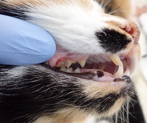
Figure 2. Examination of the right sided premolars/molars in a cat. This view also includes ability to assess the canine teeth.
I like to start by addressing one complete side of the mouth and then the other. Typically looking at buccal surface of the canine teeth and premolars/molars first before addressing the incisor region [Figure 3, 4]. I keep the mouth closed while examining, but may open the mouth slightly to get a good view of the buccal aspect of the mandibular molar(s). Many times while looking at the incisors this will cause dogs to sneeze, and in brachycephalic breeds can be challenging as you lift the upper lip it causes slight occlusion to the nares. If this is irritating to them, I try to leave this for after I have examined the rest of the teeth.
Abnormalities of the dental hard tissues to look for include: missing teeth, tooth fractures, pulp exposure, any change in opacity or discoloration, rotation, crowding, root exposure, furcation exposure. Soft tissue changes include gingivitis, gingival recession, ulceration or surface changes (masses, changes in color).
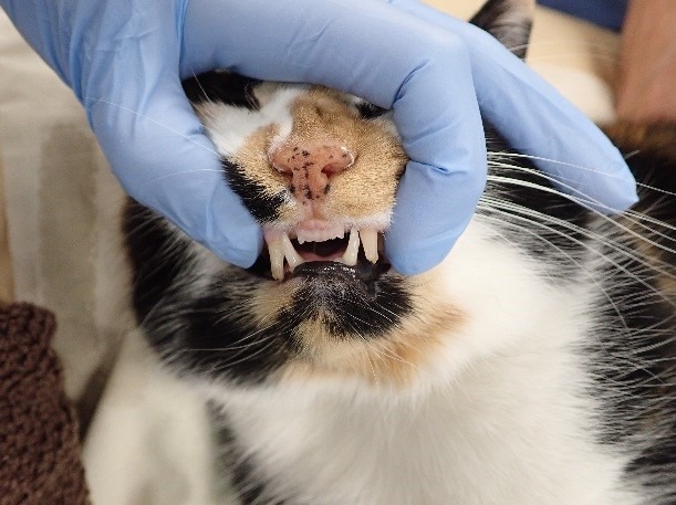
Figure 3. Examination of the canine teeth and incisor region in a cat.
Giving the mouth a generalized calculus and gingivitis index is helpful when looking back at a record and getting an idea of degree of dental disease and can also help give a rough idea when creating a treatment plan. Calculus index is graded 0-3/3 with the following guidelines: calculus index 0/3 is no appreciable calculus visible, calculus index 1 is < 1/3 of the buccal surface of the tooth covered by calculus, calculus index 2 is where 1/3 – 2/3 of the buccal surface is covered by calculus including subgingival deposition, calculus index of 3 is > 2/3 of the surface is covered by calculus and extending subgingivally. Gingival indices, similarly, are graded on a 0-3/3 scale. A gingival index of 0/3 would denote no gingivitis, gingival index of 1 is mild edema and redness, gingival index of 2 includes increased edema, redness, and (on anesthetized exam) bleeding following probing, and finally a gingival index of 3 having edema, redness, swelling, and spontaneous bleeding or bleeding with mild touch.
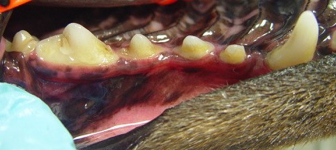
Figure 4a. Asymmetric calculus in same patient as Figure 1. Left maxillary arcade.
When grading the calculus, it is possible that different areas of the mouth may vary and while I give a generalized calculus index, I may also describe a focal area that is different from the remainder of the mouth. A very important clue can be present with asymmetric calculus accumulation. The area in which calculus is heavier will indicate the side of the pet’s mouth that they are avoiding. If they are chewing less on this side, increased plaque and calculus will accumulate. If during an examination there is such heavy calculus I cannot view the teeth, however, I can visibly see asymmetric calculus, I will bring up to the owners that I have a concern for discomfort such as a possible tooth fracture or abscessed tooth. [Figure 4]

Figure 4b. Right maxillary arcade with much heavier calculus accumulation.
Next I examine the mouth open while evaluating the molars, hard and soft palate, tongue, and sublingual area [Figure 5]. My remaining aspect of the oral exam is to look at the mucosa lining the cheeks to evaluate for buccal granulomas, masses, changes in pigmentation, or ulcerations.
The normal bite should be a scissor bite with interdigitation of the premolars and maxillary incisors should sit just slightly rostral to mandibular incisors. Other occlusal relationships (malocclusions) include: mandibular mesioclusion (maxillary brachygnathism, underbite) or mandibular distoclusion (mandibular brachygnathism, overbite), rostral or caudal crossbite, level bite and individual tooth malocclusion.
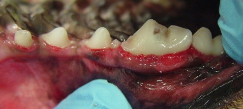
Figure 4c. Complicated crown fracture of tooth 108, same side as facial swelling and increased calculus.
When the oral exam is complete my aim is to classify and summarize the issues I have identified with the owners. Letting them know where I see “red flags” on conscious examination and teeth that likely need to be addressed as well as making them aware that additional issues may be found upon probing and viewing below the gum line with dental radiographs. Having a prepared and well educated owner will lead to the best success in treatment for each individual pet.
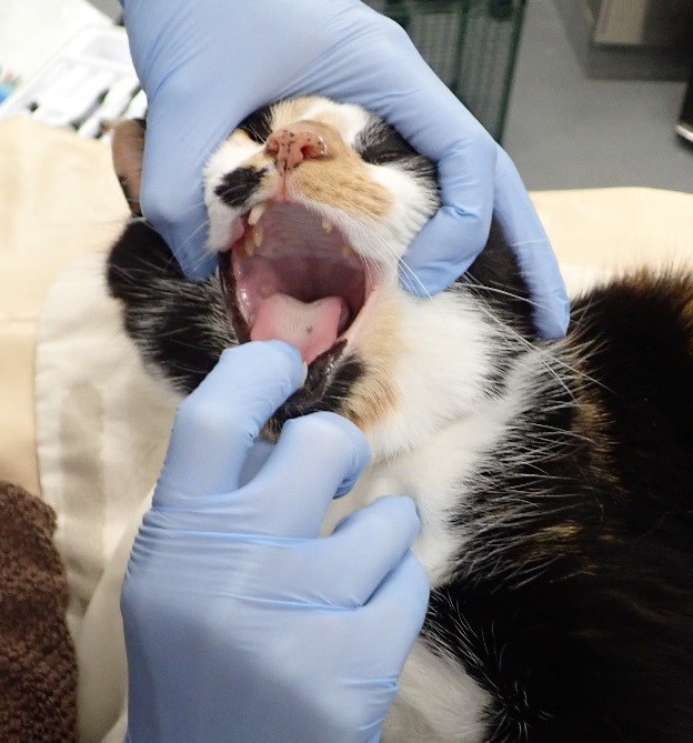
Figure 5. Positioning for caudal oral exam. The thumb of the lower hand can be used to press under the chin between the mandibles to “elevate” the tongue and visualize sublingual tissues.