-
Adopt
-
Veterinary Care
Services
Client Information
- What to Expect – Angell Boston
- Client Rights and Responsibilities
- Payments / Financial Assistance
- Pharmacy
- Client Policies
- Our Doctors
- Grief Support / Counseling
- Directions and Parking
- Helpful “How-to” Pet Care
Online Payments
Emergency: Boston
Emergency: Waltham
Poison Control Hotline
-
Programs & Resources
- Careers
-
Donate Now
 By Patrick Sullivan, DVM, DABVP (Avian practice)
MSPCA-Angell West
avianexotic@angell.org
617-989-1561
Avian respiratory emergencies can be very difficult to initially assess due to just how unstable the patient can be on presentation. Birds will often hide symptoms until they can no longer compensate and present in an advanced state of disease. This can make it impossible to complete a full physical exam without stressing the patient and causing death. The “Put It Down List” in Clinical Avian Medicine (Harrison, Lightfoot) should be consulted prior to seeing avian respiratory emergencies as it can offer potentially life-saving tips for dealing with critical patients. Some of the signs that an exam should be aborted, or held off all together are: marked dyspnea, prolonged panting, weakness/inability to stand or grasp, marked coelomic swelling, closing eyes during exam, and frank blood in the feces. Dyspneic birds should be left in their carrier if possible and placed immediately in oxygen to be monitored. A visual exam may be done at this time but all hands-on procedures should be delayed. Once the patient is more stable, an exam can be performed.
 This article is intended to provide information on common avian respiratory emergencies that may present to your practice, it is not meant to be taken as a complete overview of current literature on the topic. Please refer to the references and suggested reading list for additional information.
Supportive Care
Regardless of the cause of illness, many birds will benefit from supportive care after being admitted to the hospital. This can help to stabilize a patient, which can make them a better candidate for diagnostics and treatments.
Heat – Most sick birds, especially those that are chronically ill, may present hypothermic. For this reason, it is recommended to have heated enclosures with a humidity source in them in order to maintain an environment of 85-88℉ with a relative humidity of approximately 70%. This will reduce the amount of energy the patient needs to expend warming itself, and the humidity will aid in rehydrating mucous membranes throughout the respiratory tract.
Fluid therapy – Most critically ill birds will present with some degree of dehydration that will need to be corrected. The route with which you choose to correct this will depend on several factors and may change as treatment progresses. It is important that all parenterally administered fluids be warmed to reduce pain and aid in maintaining a normal body temperature. Oral fluid replacement may be used for mildly dehydrated patients or in conjunction with subcutaneous or intravenous administration. Subcutaneous route is the most commonly used for fluid therapy in birds. The author prefers to use the inguinal fold or the dorsum in extremely debilitated birds, taking care to avoid administering fluids directly into an air sac. In cases of marked dehydration or hypovolemia, intravenous catheter placement and fluid delivery is recommended. The most commonly accessed vessels for IV catheter placement are the basilic, or wing vein, and the medial metatarsal vein. Both sites will need to be properly protected to ensure that the patient does not chew or self-mutilate. Elizabethan collars are commonly used, as are padded bandages, to reduce this risk. In cases where venous access is not possible due to the size of the patient, or marked hypovolemia, an intraosseous catheter can be placed. These catheters are typically placed in the distal ulna or proximal tibia, and placement can be quite painful. Whenever possible, anesthesia is recommended for placement. These catheters are not typically left in place for more than 24-48 hours.
Nutritional support – Nutritional support via gavage feeding should only be done after the patient has been adequately hydrated. Several commercially available diets are available and may be chosen based on the needs of the patient. Elemental diets are often used for debilitated patients with no specific dietary restrictions due to how quickly and easily they can be broken down and used. Gavage needles can be used to deliver both nutrition and medications to patients quickly as well as to aid in hydrating the GI tract. This procedure should not be performed by inexperienced staff members as administration of food material into the respiratory tract is possible.
Clinical Signs of Respiratory Disease
Determining whether the upper or lower respiratory tract is affected can aid in determining which type of diagnostics and treatment you choose. Some types of respiratory disease begin in one part, but can quickly spread to involve the entire respiratory tract. Upper respiratory disease can manifest itself in a number of ways, but some of the more common signs are open mouth breathing, increased respiratory rate, changes in beak formation, facial and periocular swelling, and oculonasal discharge. The nares should be examined for symmetry and obstruction, as changes in shape may occur with chronic nasal plugs. Owners may also notice head shaking, rubbing the beak on things in the cage, and yawning. A change or loss of voice may indicate tracheal disease and should be investigated to rule out swelling, plaques or masses, both intra and extraluminal. Fungal diseases will often produce tracheal changes and should be considered in the presence of these symptoms. Inhalation of foreign material such as seed husks can also cause tracheal obstructions and will typically come on acutely. The choana should be examined for signs of discharge as well as blunting of the papillae. Nutritional deficiencies may lead to changes in the choanal papillae which could affect respiratory health.
Lower respiratory disease can often present with vague signs that may be confused with other diseases. Tail bobbing, open mouth breathing, coughing, exercise intolerance, general lethargy and a fluffed appearance may indicate disease. These birds are often easily stressed when handled, so physical exams may need to be shortened and oxygen therapy will need to be provided. While most causes of lower respiratory disease do not cause acute symptoms, toxin inhalation will usually present with marked dyspnea or acute death. This may be attributed to the differences of the avian respiratory tract when compared to that of mammalian. A longer exposure time to a larger surface area may cause greater effect in a very short period of time.
Common Causes
The causes of respiratory disease in birds are multiple and in many cases may be multifactorial. Below are some common causes of disease in birds. Diagnostics such as imaging, culture, PCR, and endoscopy may provide additional information on a case by case basis.
Bacterial – Several Gram negative organisms may be isolated from the respiratory tract of birds. Infection with a Gram negative pathogen will often produce copious amounts of purulent oculonasal discharge. Gram positive infections with organisms such as Streptococcus and Staphylococcus spp do occur, although these bacteria are typically considered normal flora. Chlamydia psittaci is an intracellular bacterial parasite that affects most psittacines as well as humans, causing a condition known as avian chlamydiosis in birds. This organism appears to affect different species differently, with some showing mild chronic signs and others becoming acutely ill and dying. Young animals appear to be more susceptible than older ones, although severe illness can be observed at any age. Common clinical signs include lethargy, tremors, poor plumage, conjunctivitis, dyspnea, and sinusitis. As the disease progresses, emaciation and severe dehydration may be observed. Changes in urate color (yellow to green) may be indicative of hepatic involvement. This is generally considered a poor prognostic indicator, and death may occur within one to two weeks.
Viral – Respiratory disease can be a component of many viral infections including Poxvirus, Herpesvirus, Adenovirus, Paramyxovirus and Orthomyxovirus. Avian influenza is a virus that has become quite prevalent in the past few years, with poultry outbreaks occurring in several states. The highly pathogenic avian influenza (HPAI) is differentiated from the low pathogenic strains (LPAI) by the presence of a 5 or 7 hemagglutinin. These strains of LPAI will typically occur in asymptomatic wild bird populations and then transfer to poultry. Some viruses die off, but some remain and evolve into a HPAI virus. Once this adaptation occurs, the virus rarely transfers back to the wild population. While the virus has been found in multiple species of birds, many appear to be asymptomatic carriers, and psittacine infection in general appears quite rare.
Nutritional – The most common cause of malnutrition leading to respiratory disease in companion avian species is hypovitaminosis A. Hyperkeratosis, abscessation, and blunting of the choanal papillae are typical symptoms of nutritional deficiency. These conditions are becoming less common due to commercially available complete diets, although when present they can still predispose to secondary, opportunistic infections.
Fungal – Aspergillus spp is a ubiquitous fungal organism that will often times infect patients that are immune compromised. Moldy feed, suboptimal conditions and prolonged antibiotic use may predispose animals to this condition. Affected birds may develop acute or chronic symptoms, with most acute infections leading to death. Chronic infections will often times lead to plaques throughout the respiratory tract and may also spread to organs that are in direct contact with infected air sacs. Clinical symptoms depend on the location of the lesions and the severity. Radiographs may be helpful in diagnosis, but endoscopy and biopsy will yield a more definitive diagnosis. Changes in the hemogram, especially a marked heterophilia, along with specific assays (Aspergillus Antibody, galactomannan testing) offered through universities and commercial laboratories, can be used to make a diagnosis and monitor treatment.
Another mycotic organism that can commonly infect companion species is Candida spp., which will produce white to cream colored plaques in the oropharynx that may extend down the trachea. These plaques can cause marked dyspnea and dysphagia.
Acute dyspnea – This may truly be an acute onset, or the result of a chronic, worsening condition. Tracheal obstruction is one of the more common causes of acute dyspnea and can be caused by seed inhalation or by a granuloma or aspergilloma decreasing the tracheal lumen. A high pitched squeak may be heard when the trachea is almost completely occluded, and open mouth breathing is a common symptom. Radiography may show changes in the tracheal lumen, but endoscopy will be needed to identify the obstruction. Seed husks and other inhaled food items can be stabilized by placing a needle through the trachea below the obstruction, then attempting suction to clear it. An air sac cannula will need to be placed prior to attempting these procedures. Masses outside of the trachea may be approached surgically, but the risk may be high.
Inhaled Toxin – Birds are extremely sensitive to inhaled irritants and toxins and will often times die suddenly following exposure. Others may present with irritated or inflamed mucous membranes, rhinitis, sinusitis or generally dyspneic. Common inhaled toxins include polytetrafluoroethylene gas (PTTE), which is released when Teflon pans are overheated or burned. Pans heated above 530℉ release irritating particulate matter and toxic fumes that are almost always fatal. Cigarette smoke is another common toxin that can cause coughing, sinusitis, sneezing and conjunctivitis. Owners who smoke should also wash their hands prior to handling their birds as nicotine has been shown to cause a focal dermatitis on the unfeathered skin of many birds. Household cleaners containing ammonia can cause lymphocyte derangements and general immune suppression leading to an increased risk of infection. These vapors, including bleach, can also cause mucous membrane irritation resulting in inflammation and erythema. Aerosol cleaners, fragrances or beauty products also pose a risk to birds as these may cause direct irritation to the lungs and air sacs due to the fluorocarbons and fine particulate matter that is produced. Use of these products around birds should be avoided if at all possible.
Diagnosis and Treatment
Diagnosis of any type of respiratory disease should begin with a physical exam, where you will be able to evaluate the patient and assess whether additional diagnostics will be able to be performed. Staging these tests is very common in patients with respiratory distress, and beginning with procedures that will cause little to no stress will be most beneficial. A complete blood count, serum biochemistry and cytology of the choana or trachea may be able to be performed rather quickly and can yield quite valuable information. Specific assay panels may identify pathogens such as Chlamydia psittaci or Aspergillus spp. Procedures such as a nasal flush will help in identifying pathogens through cytology and culture. Radiographs and endoscopy are potentially more invasive but may yield the location, and allow for biopsy, of specific lesions.
Treatment of respiratory disease should be based on test results when possible, but due to the unstable nature of some patients, this may not be an option. Supportive care, as mentioned above, should be integrated into most treatment plans as warranted. Systemic medications may involve antibiotic or antifungal medication based on culture, sensitivity and organism identification. Non-steroidal anti-inflammatory drugs and bronchodilators should be considered as well. Glucocorticosteroids are contraindicated for use in avian species due to marked immune suppression following administration. Emergency procedures such as intubation or air sac cannula placement may be needed and should be considered on a case by case basis.
Common Emergency Procedures
Endotracheal intubation – Intubation of most birds is quite easy due to the rostral location of the glottis and the lack of an epiglottis. Uncuffed tubes should be used for this procedure to reduce the risk of tracheal trauma and secondary stenosis. The tube should only be advanced a short distance past the opening of the glottis, as the trachea narrows drastically and could be easily traumatized.

Images 1 & 2: Endotracheal intubation of a duck – The glottis of a bird can be identified on the ventral aspect of the oral cavity. Note the lack of an epiglottis, making introduction of an endotracheal tube easier than in many mammal species. Only uncuffed endotracheal tubes should be used due to the risk of damage to the trachea, which is made up of complete cartilaginous rings, when inflating cuffed tubes. Endotracheal tubes should be introduced only 1-2cm as the trachea in some species can narrow dramatically, or bifurcate.

Image 3: An air sac cannula being used to deliver oxygen via the caudal abdominal air sac. These can be sutured in place using a finger trap technique and secured to the body.
Air sac cannula placement – This technique is only indicated for upper respiratory disease or obstruction that has not affected the lower respiratory tract. The positioning for this cannula involves right lateral recumbency with the pelvic limb pulled caudally. The area caudal to the last rib is surgically cleaned and prepped and a small skin incision is made using a scalpel. The muscle wall is bluntly penetrated using a small pair of mosquito hemostats and opened to allow the cannula to be inserted into the opening. The cannula is sutured to the skin and attached to a non-rebreathing system. The reservoir bag can be used to ensure proper cannula placement by instilling a breath into the patient and making sure the body wall rises with it.
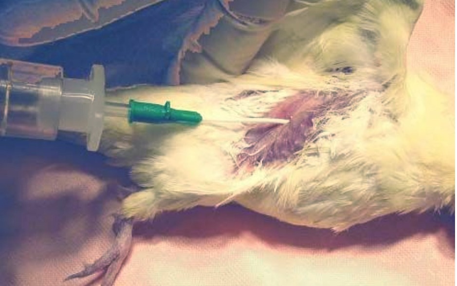
Coelomocentesis – This procedure is indicated when a bird presents with dyspnea secondary to coelomic effusion. It is recommended to not lay the patient on its side, but rather to keep it in an upright position. The ventral coelom is plucked and surgically prepped. A 22g IV catheter is advanced at a 90° to the body wall until fluid is seen in the hub. The catheter sheath is advanced, the stylet is removed, and an extension set with syringe is attached to the catheter. The fluid should be removed using the minimal amount of pressure needed to reduce the risk of damage to any internal structures. Patients should be recovered in oxygen and monitored for continued dyspnea.
References and Suggested Reading
- Ritchie, Harrison, Harrison. Avian Medicine: Principles And Applications . Wingers Publishing, Inc. 1994
- Speer, Brian. Current Therapy In Avian Medicine And Surgery . Elsevier Publishing. 2016
- Harrison, Lightfoot. Clinical Avian Medicine . Spix Publishing, Inc. 2006
- King, McLelland. Birds-Their Structure And Function . Bailliere Tindall. 1984