-
Adopt
-
Veterinary Care
Services
Client Information
- What to Expect – Angell Boston
- Client Rights and Responsibilities
- Payments / Financial Assistance
- Pharmacy
- Client Policies
- Our Doctors
- Grief Support / Counseling
- Directions and Parking
- Helpful “How-to” Pet Care
Online Payments
Referrals
- Referral Forms/Contact
- Direct Connect
- Referring Veterinarian Portal
- Clinical Articles
- Partners in Care Newsletter
CE, Internships & Alumni Info
CE Seminar Schedule
Emergency: Boston
Emergency: Waltham
Poison Control Hotline
-
Programs & Resources
- Careers
-
Donate Now
 Sue Casale, DVM, DACVS
Angell Surgery Service
617-541-5048
surgery@angell.org
Forelimb lameness is one of the most frustrating conditions to diagnose and treat. Often dogs will have mild and intermittent lameness with little to no reaction on orthopedic examination. Numerous conditions exist which result in forelimb lameness, so often times many diagnostics are needed to determine the cause, and despite multiple diagnostics a cause may not be immediately found. This results in frustration on the part of both the owner and the veterinarian. In addition, pathology in multiple joints is common and determining the culprit of the lameness may be difficult. Studies have shown that shoulder pathology is more likely to cause lameness than elbow pathology.1 History may lend some clues to the origin of the lameness. Lameness from shoulder pathology tends to be weight bearing and consistent with an insidious onset while elbow lameness is more acute and episodic.

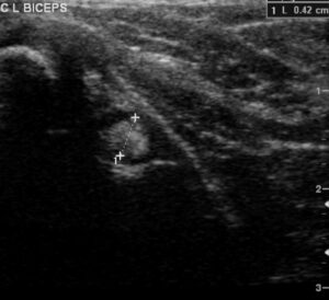
Ultrasound image of biceps tendon
One common cause of shoulder lameness is biceps tenosynovitis. This condition is often seen in middle-age-to-older, medium-to-large breed dogs with the Labrador Retriever and Rottweiler the most frequent breeds affected. The biceps brachii muscle originates on the supraglenoid tubercle of the scapula and inserts on the radius and ulna. The biceps tendon crosses the joint and is both a dynamic and static stabilizer of the shoulder joint.2 Strain injury to the tendon from overuse or trauma, both direct and indirect, can result in inflammation of the tendon and surrounding sheath. Pain is often elicited on direct biceps tendon palpation while the shoulder is flexed and the elbow extended although general shoulder pain may also elicit a response.3 Shoulder radiographs are necessary to rule out other pathology and may show mineralization within the biceps groove, however, normal radiographs do not rule out biceps tenosynovitis.1,4 Contrast arthrography is generally low yield, but may show a filling defect in the biceps groove if there are fibrous adhesions present. Ultrasound is the modality of choice to diagnose biceps disease.5 Other soft tissue structures can be examined and the contralateral biceps tendon can be used for comparison. Ultrasound of the biceps tendon when tenosynovitis is present may reveal an enlarged, hypoechoic tendon with fiber pattern disruption. A lucent line around the tendon indicating fluid within the sheath may be seen and an irregularity to the bicipital groove from osteophytes is seen in chronic cases.

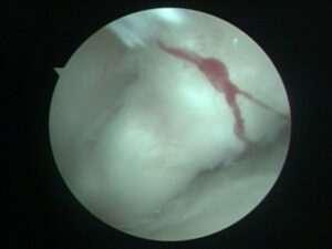
Arthroscopic view of biceps tendon
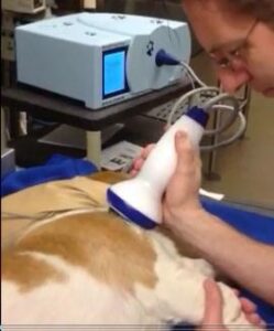
Treatment of biceps tenosynovitis starts with six weeks of strict rest and non-steroidal anti-inflammatory medication. The joint/tendon sheath can be injected with a steroid such as Depo-Medrol. Direct injection into the tendon is not recommended as it may predispose patients to tendon rupture.6 Medical management can be successful in more than 50% of cases, but return to normal activity too early can disrupt the healing, slow the recovery and cause chronic lameness.3,7 If there is no improvement after medical management, or if the lameness returns, surgical treatment may be required. Extracorporeal shockwave therapy (ESWT) is a new modality to treat tendon injuries. With ESWT, high energy acoustic pressure waves are delivered to the tendon. Although the mechanism of action is not completely understood, several hypotheses exist to explain its healing mechanism.8 Neo-angiogenesis, modulation of nitric oxide, suppression of NF-kappaB, and increased expression of TGF-beta1 and IGF-1 are all thought to play a role. ESWT decreases inflammation and speeds the healing process through increased release of angiogenic growth factors, disintegration of calcifications within the tendon, and increased tensile strength within the tendon by improving fiber alignment.9 ESWT is also thought to recruit stem cells to aid in repair and increases the cellular release of bone morphogenic protein. Sedation is required for treatment with ESWT because of mild discomfort during the procedure and the general recommendation is three treatments, three weeks apart. Up to 85% of dogs will show improvement with ESWT. If improvement is incomplete or temporary, surgery can be considered. Surgical transection of the tendon eliminates the repetitive trauma from the tendon stretching across the shoulder joint. The transected end of the tendon can be surgically attached to the proximal humerus with a bone screw (biceps tenodesis) or can be left to form adhesions to the humerus over time (natural tenodesis). Arthroscopy allows direct visualization of the tendon and its origin and allows transection of the tendon with minimal tissue trauma. Arthroscopic tenotomy results in excellent outcome in most dogs with many experiencing immediate pain relief.10,11 Mean time to full recovery is three weeks.

Extracorporeal Shockwave Therapy (ESWT)
Although biceps tenosynovitis can be frustrating to diagnose, ultrasound is an excellent modality to help diagnose shoulder lameness from biceps disease. Tenosynovitis has a high response rate to medical management and ESWT, however, surgery is a good option for patients that do not respond with good to excellent results in most dogs.
For more information about Angell’s Surgery service, please visit www.angell.org/surgery. To contact Angell’s surgeons by phone or to refer a patient to the Angell Surgery service, please call 617 541-5048 or e-mail surgery@angell.org. You can also reach Dr. Casale at scasale@angell.org.
References
1Cogar DM, Cook CR, Curry SL et al. Prospective evaluation fo techniques for differentiation shoulder pathology as a source of forelimb lameness in medium and large breed dogs. Vet Surg 2008;37:132-141.
2Sidaway, BK, McLaughlin RM, Elder SH et al. Role of the tendons of the biceps brachii and infraspinatus muscles and the medial genohumeral ligament in the maintenance of passive shoulder joint stability in dogs. Am J Vet Res 2004;65:1216-1222.
3Stobie D, Wallace LJ et al. Chronic bicipital tenosynovitis in dogs: 29 cases (1985-1992). J Am Vet Med Assoc. 1995;207:201-207.
4Akerblom S, Sjostrom L. Evaluation of clinical, radiographical and cytologic findings compared to arthroscopic findings in shoulder lameness in the dog. Vet Comp Orthop Traumatol 2007;20:136-141.
5Kramer M, Gerwing M, Sheppard, C, Schimke, E. Ultrasonography for the diagnosis of disease of the tendon and tendon sheath of the biceps brachii muscle. Vet Surg 2001;30:64-71.
6Halpern AA, Horowitz BG, Nagel DA. Tendon ruptures associated with corticosteroid therapy. West J Med. 1977;127:378-382.
7Bruce WJ, Burbidge HM, Broome CJ. Bicipital tendinitis and tenosynovitis in the dog: a study of 15 cases. N Z Vet J 2000;48:44-52.
8Shrivastava, SK, and Kailash. Shock wave treatment in medicine. J Biosci 2005;30:269-275.
9Farr S, Sevelda F, et al. Extracorporeal shockwave therapy in calcifying tendinits of the shoulder. Knee Surg Sports Traumatol Arthrosc 2011;19:2085-2089.
10Wall CR, and Taylor R Arthroscopic biceps brachii tenotomy as a treatment for canine bicipital tenosynovitis. J An Anim Hosp Assoc 2002;38:169-175
11Bergenhuyzen ALR, Vermote KAG, van Bree H, and Van Ryssen B. Long-term follow-up after arthroscopic tenotomy for partial rupture of the biceps brachii tendon. Vet Comp Ortho Traumatol 2010;23:51-55.