-
Adopt
-
Veterinary Care
Services
Client Information
- What to Expect – Angell Boston
- Client Rights and Responsibilities
- Payments / Financial Assistance
- Pharmacy
- Client Policies
- Our Doctors
- Grief Support / Counseling
- Directions and Parking
- Helpful “How-to” Pet Care
Online Payments
Emergency: Boston
Emergency: Waltham
Poison Control Hotline
-
Programs & Resources
- Careers
-
Donate Now
 By Pamela Mouser, DVM, MS, DACVP
![]()
angell.org/lab
Oral masses, especially from dogs, are a common submission to Angell’s biopsy service. This may be in part due to clinical signs alerting the owner to the presence of an oral mass, such as drooling, oral bleeding, halitosis, or anorexia, leading to veterinary evaluation of affected dogs. A subset of oral masses may also be detected on physical examination during a wellness visit or during veterinary assessment for another condition. At Angell, the dentistry service is a large contributor of oral masses. Veterinary dentistry has progressed beyond “dental cleaning” or “dental prophylaxis” to encompass a much broader and more detailed orofacial evaluation termed Comprehensive Oral Health Assessment and Treatment (COHAT). Since this examination is performed annually (sometimes more often, depending on the patient) under general anesthesia, it has the added benefit of providing early detection of oral masses, which can then be sampled or excised during the procedure. This article retrospectively summarizes data from canine oral masses submitted to Angell Pathology over a period of approximately five years, and compares Angell submissions to published reports.
Study Design
Surgical biopsies submitted to Angell Pathology from October 2014 to January 2020 were searched to identify any samples designated as “oral cavity,” “maxilla bone,” or “mandible bone.” The designation of “oral cavity” encompassed typical intraoral structures including gingiva, tongue, tooth, palate, cheek, etc. The data set was limited to canine patients. Lesions not considered masses or mass-like in nature (including inflammation, ulceration, or granulation tissue) were removed from the data. Duplicate patient entries were evaluated to exclude multiple instances of the same lesion, such as repeat biopsy or re-excision of the same mass. Data were summarized and descriptive statistics were applied.
Results
The data set included 673 dogs with 942 masses. Of these, 522 dogs (78%) had a single mass and 151 (22%) had multiple masses. The number of masses in dogs with multiple lesions ranged from two to ten; some dogs with multiple lesions had masses submitted on more than one occasion. Dogs ranged from 0.6 – 16 years old, with a mean and median of 9 years old. There were 391 males (58.1%) of which 347 (51.6% of total population) were designated as castrated, and 282 females (41.9%) with 278 spayed (41.3%). Over 100 dog breeds were represented. Labrador retrievers (or Labrador retriever crosses) accounted for 80 (12%) of the patients, followed by 51 golden retrievers/crosses (7.6%) and 38 boxers/crosses (6%). The most common breeds are listed in Table 1.
Table 1. Most common breeds in submissions of canine oral masses from October 2014 to January 2020.
| Breed | Number |
| Labrador retriever/cross |
80 |
| Golden retriever/cross |
51 |
| Boxer/cross |
38 |
| Pit Bull/cross |
28 |
| German shepherd dog/cross |
20 |
| Maltese/cross |
19 |
| Terrier/cross |
18 |
| Yorkshire terrier/cross |
18 |
| Mixed Breed |
16 |
| Cocker spaniel/cross |
15 |
| Pug/cross |
14 |
| Shih tzu/cross |
13 |
| Boston terrier |
13 |
| Beagle/cross |
12 |
| Bulldog (English/American/cross) |
12 |
| Chihuahua/cross |
12 |
| Dachshund/cross |
12 |
| Schnauzer/cross |
12 |
| Bernese Mountain Dog |
11 |
| Standard poddle |
9 |
| Shetland sheepdog |
9 |
| French bulldog |
8 |
| Jack Russell terrier |
8 |
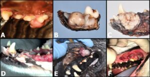
Non-neoplastic and benign diagnoses accounted for 812 (86%) of the 942 masses. The most common diagnosis was gingival fibrous hyperplasia (412 masses or 44%), followed by peripheral odontogenic fibroma (223 masses or 24%). The most common malignant diagnoses were squamous cell carcinoma and melanoma (33 masses or 3.5% each), as well as sarcoma (31 masses or 3.3% of total masses). A summary of histopathologic diagnoses is included in Table 2. In addition, Figure 1 depicts various presentations of oral masses in dogs biopsied at Angell. Note that the clinical manifestation of masses may overlap, making it difficult to impossible to distinguish between some non-neoplastic, benign, and malignant entities based on gross appearance alone.
Table 2. Histopathologic diagnosis of canine oral masses from October 2014 to January 2020.
| Histopathologic Diagnosis |
Number of Masses |
| Non-neoplastic diagnoses |
486 (51.6%) |
|
412 (43.7%) |
|
42 (4.5%) |
|
16 (1.7%) |
|
7 (0.7%) |
|
3 (0.3%) |
|
3 (0.3%) |
|
2 (0.2%) |
|
1 (0.1%) |
| Benign neoplasms |
326 (34.6%) |
|
223 (23.7%) |
|
49 (5.2%) |
|
20 (2.1%) |
|
13 (1.4%) |
|
7 (0.7%) |
|
7 (0.7%) |
|
5 (0.5%) |
|
2 (0.2%) |
| Malignant neoplasms |
130 (13.8%) |
|
33 (3.5%) |
|
33 (3.5%) |
|
31 (3.3%) |
|
7 (0.7%) |
|
7 (0.7%) |
|
5 (0.5%) |
|
3 (0.3%) |
|
3 (0.3%) |
|
2 (0.2%) |
|
1 (0.1%) |
|
1 (0.1%) |
|
1 (0.1%) |
|
1 (0.1%) |
|
1 (0.1%) |
|
1 (0.1%) |
Figure 1. Clinical and gross photographs of canine oral masses submitted for histopathology at Angell. A: A pink smooth firm raised mass of the palatal gingiva diagnosed as peripheral odontogenic fibroma. B: A formalin-fixed, partial mandibulectomy sample with a smooth nodular mass bearing some resemblance to image A, but compatible with a sarcoma on histopathology. C: A formalin-fixed rostral mandibulectomy sample showing a rough, pale tan to white verrucous gingival mass diagnosed as acanthomatous ameloblastoma. D: A gross image from Angell archives showing irregular, coalescing gingival expansion and hyperemia, corresponding to T-cell lymphoma. E: A multinodular pink-red mass on the buccal mucosa of a dog with disseminated osteosarcoma. The oral mass was an amelanotic melanoma. F: An ulcerated, irregular plaque-like mass of the upper gingiva corresponding to squamous cell carcinoma.
Discussion
Canine oral masses submitted to Angell Pathology over the specified time period were predominantly non-neoplastic or benign. It is important to note that Angell submissions are potentially skewed to include a greater number of small or incidental lesions detected and removed at the time of COHAT. Similar lesions may be missed during physical examination of a non-anesthetized patient or, if detected, cannot be immediately sampled and therefore biopsy may be delayed or not pursued in the absence of clinical signs. Similar numbers of squamous cell carcinoma, melanoma, and sarcoma were diagnosed in the malignant category of this population. It is possible that some oral masses diagnosed as sarcoma represent a spindloid variant of amelanotic melanoma, but were not further characterized due to lack of ancillary testing. Thus, application of immunohistochemistry to cases of oral sarcoma may have increased the total number of melanoma diagnoses (and decreased sarcoma cases). The following paragraphs highlight statistics from prior reports on canine oral masses, in comparison with Angell data.
A 2019 retrospective study by Mikiewicz et al documented 486 cases of oral masses in dogs and cats. Similar to the data shown above, there was a predominance of benign and hyperplastic lesions in dogs, although composing a much lower overall percent of cases (53.2%) when compared to Angell submissions (86%). The difference in proportions between the two data sets may be related to increased detection/submission of small or “incidental” gingival masses in the Angell population, as described above; the inclusion of multiple masses from individual Angell dogs; and also because the 2019 retrospective study included inflammatory lesions without which the percentage of benign/hyperplastic lesions would have been 62.4%. The most common malignant diagnoses in the 2019 study were melanoma followed by squamous cell carcinoma and fibrosarcoma, similar to the top three malignancies seen by Angell Pathology.
A 2018 retrospective study of canine and feline oral biopsies from a dental specialty practice reported 62% of the 403 canine lesions were classified as either inflammatory or odontogenic. The study did not specify how hyperplastic lesions were categorized. Squamous cell carcinoma, fibrosarcoma, and melanoma (with equal numbers of osteosarcoma) were the most common malignant diagnoses in descending order. The 2018 study included plasma cell tumor in the “malignant” category, while this tumor was considered benign in the Angell data and in the above 2019 retrospective study.
A 2010 retrospective study of 280 oral biopsies in Swedish dogs reported a predominance of reactive or benign diagnoses (66% of cases), with gingival hyperplasia (24%) and peripheral odontogenic fibroma (21%) most common. Melanoma, sarcoma, and squamous cell carcinoma were the most common malignant diagnoses in descending order.
A retrospective summary of canine oral biopsies from the University of California-Davis Dentistry and Oral Surgery Service had 75% of all diagnoses categorized as benign or non-neoplastic (including inflammation and hyperplasia). Melanoma, squamous cell carcinoma, and fibrosarcoma were the most common malignancies reported.
The summarized data from canine oral biopsies submitted to Angell Pathology over a five year period is similar to retrospective studies, specifically the predominance of benign/non-neoplastic lesions and the most prevalent malignancies. Interestingly, the proportion of benign or non-neoplastic lesions is highest in the Angell group, likely related to selection bias (the types of cases submitted to Angell Pathology). The proportion of benign/non-neoplastic diagnoses in the Angell population would have been even higher if inflammatory lesions had been included. In general, canine oral masses are more often benign or non-neoplastic. When malignant, the most common lesions include squamous cell carcinoma, melanoma, and sarcoma.
References
- Mikiewicz M, Pazdzior-Czapula K, Gesek M, Lemishevskyi V, Otrocka-Domagala I. Canine and feline oral cavity tumours and tumour-like lesions: a retrospective study of 486 cases (2015-2017). J Comp Path 2019;172:80-87.
- Regezi JA, Verstraete FJM. Clinical-pathologic correlations. In: Verstraete FJM, Lommer MI, Arzi B, eds. Oral and Maxillofacial Surgery in Dogs and Cats. 2nd ed. St. Louis, MO: Elsevier; 2020:424.
- Svendenius L, Warfvinge G. Oral pathology in Swedish dogs: a retrospective study of 280 biopsies. J Vet Dent 2010;27:91-97.
- Wingo K. Histopathologic diagnoses from biopsies of the oral cavity in 403 dogs and 73 cats. J Vet Dent 2018;35:7-17.
