-
Adopt
-
Veterinary Care
Services
Client Information
- What to Expect – Angell Boston
- Client Rights and Responsibilities
- Payments / Financial Assistance
- Pharmacy
- Client Policies
- Our Doctors
- Grief Support / Counseling
- Directions and Parking
- Helpful “How-to” Pet Care
Online Payments
Referrals
- Referral Forms/Contact
- Direct Connect
- Referring Veterinarian Portal
- Clinical Articles
- Partners in Care Newsletter
CE, Internships & Alumni Info
CE Seminar Schedule
Emergency: Boston
Emergency: Waltham
Poison Control Hotline
-
Programs & Resources
- Careers
-
Donate Now
 By Patty Ewing, DVM, MS, DACVP
(anatomic and clinical pathology)
angell.org/lab
617-541-5014
Introduction
Cytologic evaluation of cutaneous and subcutaneous mass aspirates is a convenient, rapid way of obtaining a definitive or presumptive diagnosis in a majority of patients. In one study comparing cytologic and histopathologic test results of cutaneous and subcutaneous lesions in 243 specimens, the diagnosis was in agreement in 90.9% of cases.1 The results of cytologic evaluation may provide information about prognosis, guidance for next diagnostic steps and/or treatment. Sedation or anesthesia is rarely needed for sample collection. Samples can often be collected, prepared and evaluated microscopically in a matter of minutes. The category of discrete round cell tumors is composed primarily of cells of the hemolymphatic system. In addition to the round to oval shape of the cells, the distinguishing morphologic feature is their discrete nature that lacks cellular junctions resulting in singly occurring cells rather than cohesive aggregates (epithelial tumors) or loose aggregates of cells associated with extracellular matrix (mesenchymal/spindle cell tumors).2 Aspirates of round cell tumors generally yield a large number of neoplastic cells making them rewarding to evaluate. In this color atlas, cytologic characteristics of the following six canine round cell tumors will be highlighted: canine cutaneous histiocytoma, histiocytic sarcoma, mast cell tumor, lymphoma, plasma cell tumor, and transmissible venereal tumor (TVT). Table 1 provides a summary of cytologic features and typical locations for each round cell tumor type.
Table 1. Cytologic Characteristics of Cutaneous/Subcutaneous Discrete Round Cell Tumors
| Type | Common Appearance | Cytologic Characteristics2 |
| Histiocytoma | Single smooth pink raised hairless mass; often on head, pinnae | Relatively uniform medium round cells w/moderate-sized, round to slightly indented nucleus, finely etched chromatin, moderate amount of pale, slightly granular cytoplasm; few mitotic figures |
| Histiocytic sarcoma | Single or multiple, purple/red nodules; often with visceral involvement or periarticular location | Large round to spindle cells with one or more large round nuclei, marked pleomorphism, abundant pale cytoplasm often vacuolated or exhibiting phagocytosis of RBCs or WBCs; mitotic figures may be present |
| Mast cell tumor | Single or less often multiple white to light yellow or hemorrhagic masses or plaques; ulceration common; visceral involvement possible | Medium round cells with central round nucleus often obscured by numerous fine to course, purple cytoplasmic granules filling moderately abundant pale cytoplasm |
| Lymphoma | Multiple off white or red to purple nodules in nonepitheliotropic type | Medium to large round cells with high nucleus-to-cytoplasm (N:C) ratio, finely granular chromatin, prominent nucleoli, scant medium blue cytoplasm; mitotic figures common in high grade lymphoma |
| Plasma cell tumor | Single raised pink nodule on head, trunk or limbs; usually not associated w/multiple myeloma | Medium round cells with a large round eccentric nucleus, coarsely reticulated to stippled chromatin, + indistinct nucleoli, abundant medium to deep blue cytoplasm often with perinuclear clear zone; mitotic figures may be present. |
| Transmissible Venereal Tumor | Single or more often multiple nodular pedunculated to cauliflower-like masses on external genitalia of sexually active dogs | Large round cells with moderate-sized, round nuclei, coarsely stippled chromatin, small to prominent nucleoli, moderate amount of pale cytoplasm with few small distinct cytoplasmic vacuoles; mitotic figures may be present. |
Canine Cutaneous Histiocytoma
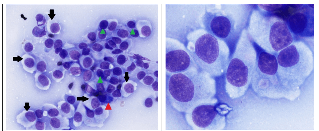
Histiocytomas are benign tumors composed of Langerhans cells, which are dendritic, antigen-presenting cells of the skin. Histiocytoma occurs most commonly as a solitary skin mass in dogs under 2 years of age, but can occur in dogs of any age.3 Scottish terriers, bull terriers, boxers, English cocker spaniels, flat coated retrievers, Doberman pinschers and Shetland sheepdogs are predisposed toward development of histiocytomas.4 The majority of tumors undergo spontaneous regression due to immunologic reactivity.4 Cytologic appearance is shown in Figure 1.

Figure 1. Aspirate from a cutaneous histiocytoma. Left. Note large discrete round to oval cells (black arrows) of relatively uniform size that have a large, round to oval, slightly eccentric nucleus and moderate amount of cytoplasm. Low numbers of small lymphocytes (green arrow heads) and a plasma cell (red arrow head) are present. (Diff-Quik, 600x magnification.) Right. Higher magnification of histiocytoma cells showing finely etched chromatin with 1-3 small indistinct nucleoli in the nucleus and abundant, slightly granular, pale blue cytoplasm. (Diff-Quik, 1500x magnification.)
Histiocytic Sarcoma (Malignant Histiocytosis)
Histiocytic sarcoma may occur as localized masses in the subcutaneous tissue or periarticular region or as disseminated disease (lymph nodes, bone marrow, spleen, liver, lung, skin). The disseminated form is commonly referred to as malignant histiocytosis. Localized histiocytic sarcomas occur most commonly in middle-aged or older flat-coated retrievers, golden and Labrador retrievers and rottweilers.5 Malignant histiocytosis has a predilection for Bernese mountain dogs as well as the aforementioned breeds.5 The disease is uniformly fatal after a progressive disease course. Cytologic appearance is shown in Figure 2.

Figure 2. Aspirates of histiocytic sarcoma. Left. Note large discrete singly occurring round to oval cells that have a large, round, eccentric nucleus, course chromatin, multiple round nucleoli and moderate amount of vacuolated cytoplasm containing darkly stained hemosiderin pigment, phagocytized erythrocytes or leukocytes (black arrows). The green arrow identifies a neoplastic cell with a bizarre mitotic figure. Red arrow heads identify neutrophils for size comparison. (Diff-Quik, 1000x magnification.) Right. Histiocytic sarcoma with both round and pyriform to spindle-shaped cells (black arrows) arranged in non-cohesive aggregates. Note multinucleation of some of the cells. (Diff-Quik, 500x magnification.)
Mast Cell Tumor
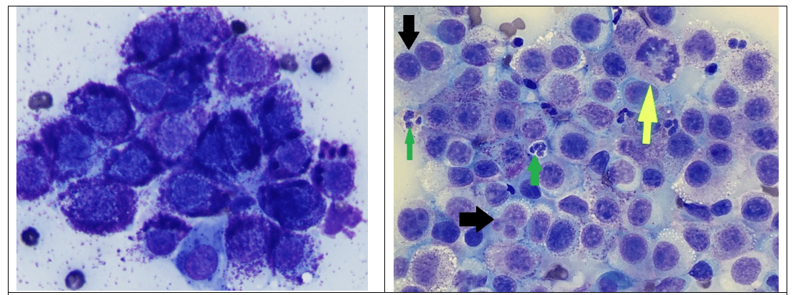
Mast cell tumors are one of the most rewarding round cell tumors to diagnose because they are readily identified by the presence of their distinctive purple mast cell tumors. Multiple dog breeds are predisposed to developing mast cell tumors, which may be solitary or multicentric. Most tumors occur in middle-aged and older dogs. A cytologic grading scheme for mast cell tumors was recently proposed and may be helpful in differentiating low grade and high grade tumors, although histologic evaluation remains the gold standard for mast cell tumor grading. In the study that evaluated this grading scheme, dogs with low grade tumors had a prolonged survival relative to dogs with high grade tumors (mean survival time of 364.6 + 42.5 days with high grade tumors). Dogs with cytologic high grade mast cell tumors were 25 times more likely to die within the 2-year follow-up period than dogs with low grade tumors. The grading scheme had 88% sensitivity and 94% specificity relative to the two-tiered histology grading system.6 Cytologic appearance of a low grade versus a high grade tumor is shown in Figure 3.

Figure 3. Aspirate of low grade (left) and high grade (right) mast cell tumors. Left. Low grade mast cell tumor has medium discrete round to oval cells (black arrows) of relatively uniform size with a medium, round central nucleus partially observed by numerous small purple granules that fill the abundant cytoplasm. (Diff-Quik, 1000x magnification.) Right. High grade mast cell tumor has sparsely granulated discrete round cells exhibiting multinucleation (black arrows), bizarre mitotic figure (yellow arrow), and nuclear atypia featuring multiple prominent nucleoli. Green arrows identify neutrophils and eosinophils for size comparison. (Diff-Quik, 600x magnification.)
Lymphoma
Cutaneous lymphoma exists in two different forms: 1) epitheliotropic, which is typically small to intermediate cell size, and 2) nonepitheliotropic, which is typically of intermediate to large cell size. Epitheliotropic lymphoma will not be covered here since this entity requires histopathology to identify epitheliotropism for making the diagnosis. Briards, English cocker spaniels, bulldogs, boxers, Scottish terriers and golden retrievers are predisposed to cutaneous lymphoma.5 Dogs with nonepitheliotropic lymphoma have single or more often multiple cutaneous nodules. The disease is progressive with eventual involvement of lymph nodes and/or viscera. Cytologic appearance of nonepitheliotropic lymphoma compared to cutaneous plasmacytoma is shown in Figure 4.
Plasmacytoma (Plasma Cell tumor)
Plasmacytomas typically arise de novo from cutaneous plasma cells and are not associated with bone marrow involvement. Multiple myeloma (plasma cell neoplasia in the bone marrow and other organs) can be associated with skin involvement, but this occurs very infrequently.4 Older dogs are more commonly affected with cutaneous plasmacytomas. Cocker spaniels, Airedale terriers, Kerry blue terriers, standard poodles, Yorkshire terriers, and Scottish terriers are predisposed.5 Most cutaneous plasmacytomas are cured with complete surgical excision. Cytologic appearance of plasmacytoma compared to lymphoma is shown in Figure 4.

Figure 4. Cytologic appearance of lymphoma (left) compared to plasmacytoma (right). Left. Note lymphoma cells are medium to large and have a higher N:C ratio and more finely stippled chromatin compared to the plasmacytoma. Yellow arrows identify non-neoplastic small lymphocytes for size comparison. The green arrow identifies a bizarre mitotic figure. (Diff-Quik, 750x magnification.) Right. Neoplastic plasma cells have increased amounts of deep blue cytoplasm compared to lymphoma with a more eccentrically placed nucleus that has a coarser or reticular chromatin pattern. Binucleation (black arrow) or multinucleation and prominent anisokaryosis (green arrows identify neoplastic cells with larger nuclei) are common features. Both lymphoma and plasmacytoma may display perinuclear clear zones and mitotic figures. (Diff-Quik, 750x magnification.)
Transmissible Venereal Tumor (TVT)
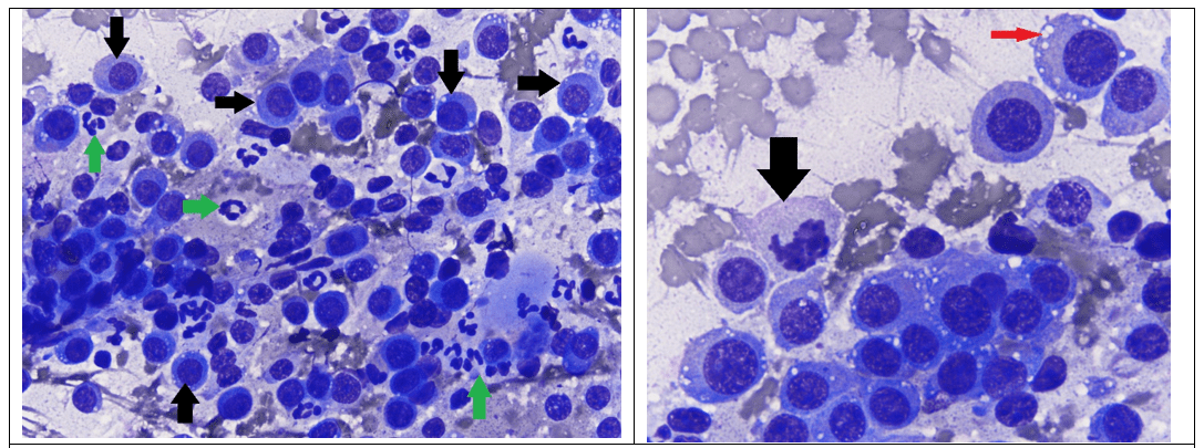
The cell of origin of TVT remains a mystery and it is unique among the round cell tumors in being transmitted by physical transplantation in sexually active dogs. The chromosome count is 59 rather than the normal 78 found in other cell types.4 In addition to external genitalia, masses may be found on the lips and other portions of the skin or mucosa that come into contact with the genitalia. Tumors initially grow rapidly, then become static for a time, and may eventually undergo spontaneous remission several months later. TVT infrequently metastasizes to regional lymph nodes and rarely, to viscera.4 Treatment with vincristine is highly effective at achieving complete remission in most dogs. Cytologic appearance of TVT is shown in Figure 5.

Figure 5. Cytologic appearance of transmissible venereal tumor. Left. Note large discrete round cells with moderate-sized, round, slightly eccentric nuclei and a moderate amount of pale blue cytoplasm. Green arrows identify neutrophils for size comparison. (Diff-Quik, 600x magnification.) Right. Higher magnification of neoplastic cells showing coarsely stippled chromatin, one or more small to medium-sized dark nucleoli, cytoplasmic vacuoles (thin red arrow) and a mitotic figure (black arrow). (Diff-Quik, 1200x magnification.)
Summary
Cytologic examination of cutaneous and subcutaneous round cell tumors is often rewarding due to the high cell yield on fine-needle aspiration. With practice, differentiation of the six distinct types of cutaneous round cells shown here can be readily achieved in the vast majority of cases. The results of cytologic evaluation may provide information about prognosis, guide next diagnostic steps or allow for rapid treatment.
References
- Ghisleni G, Roccabianca P, Ceruti R, et al: Correlation between fine-needle aspiration cytology and histopathology in the evaluation of cutaneous and subcutaneous masses from dogs and cats. Vet Clin Pathol 35(1):24-30, 2006.
- Valenciano, A and Cowell R: Cowell and Tyler’s Diagnostic Cytology and Hematology of the Dog and Cat, 4th St. Louis, MO: Elsevier, 2014, p. 70, 93-98.
- Scott DW, Miller Jr WH: Griffin CE: Muller & Kirk’s small animal dermatology, 6th Philadelphia, PA: Saunders, 2001.
- Meuten D, Editor: Tumors in Domestic Animals, 4th Ames, Iowa: Iowa State Press, 2002, pp. 109, 113, 115-117, 163.
- Meuten D, Editor: Tumors in Domestic Animals, 5th Wiley Blackwell, 2016, pp. 171, 172, 328.
- Camus M, Priest H, Koehler J, et al: Cytologic criteria for mast cell tumor grading in dogs with evaluation of clinical outcome. Vet Pathol 53(6): 1117-1123, 2016.