-
Adopt
-
Veterinary Care
Services
Client Information
- What to Expect – Angell Boston
- Client Rights and Responsibilities
- Payments / Financial Assistance
- Pharmacy
- Client Policies
- Our Doctors
- Grief Support / Counseling
- Directions and Parking
- Helpful “How-to” Pet Care
Online Payments
Emergency: Boston
Emergency: Waltham
Poison Control Hotline
-
Programs & Resources
- Careers
-
Donate Now
 By Patty Ewing, DVM, MS, DACVP
Anatomic and Clinical Pathologist
angell.org/lab
617-541-5014
Introduction
Cytologic evaluation of feline cutaneous mass aspirates is a convenient, rapid way of obtaining a definitive or presumptive diagnosis in a majority of patients. In one study comparing cytologic and histopathologic test results of cutaneous and subcutaneous lesions in 243 specimens from cats and dogs, the diagnosis was in agreement in 90.9% of cases.1 The results of cytologic evaluation may provide information about prognosis, guidance for next diagnostic steps and/or treatment. Sedation or anesthesia is rarely needed for sample collection. Samples can often be collected, prepared and evaluated microscopically in a matter of minutes.
In a retrospective study of more than 9,000 feline cutaneous tumors, 6.6% were non-neoplastic and 93.4% were neoplastic, of which 52.7% were considered malignant. The ten most common skin tumor types accounted for 80.7% of cases, with the four most common being basilar epithelial tumor, fibrosarcoma, squamous cell carcinoma and mast cell tumor.2 In this color atlas, cytologic characteristics of the four most common skin tumors will be reviewed. Table 1 provides a summary of cytologic features and typical locations for each round cell tumor type.
Table 1. Cytologic Characteristics of Cutaneous/Subcutaneous Discrete Round Cell Tumors
| Type | Common Appearance | Cytologic Characteristics2 |
| Basilar epithelial tumor |
Solitary firm raised pigmented hairless mass; often on head or neck |
Cohesive aggregates of small basaloid epithelial cells with high nuclear-to-cytoplasmic ratios, scant blue cytoplasm and monomorphic round to oval nuclei. Melanin pigment may be present within the cells or in background with cystic amorphous blue material and degenerate cells. |
| Squamous cell carcinoma |
Solitary or multiple, plaque-like or raised proliferative or ulcerative masses; often on pinnae or face. | Medium to large, oval/round to angular epithelial cells occur singly and in sheets. Cells exhibit a high nuclear-to-cytoplasmic ratio, mild to marked anisocytosis and anisokaryosis, and small condensed or larger oval vesicular nuclei with prominent nucleoli, +/- prominent perinuclear vacuolation. Concurrent neutrophilic inflammation which may be septic is common. |
| Mast cell tumor | Solitary or multiple well-circumscribed dermal masses often on head, neck and limbs. | Singly occurring, medium round cells with central round nucleus often obscured by numerous fine to course, purple cytoplasmic granules filling moderately abundant pale cytoplasm. Some stained with Diff-Quik® have non-staining granules. Some variants exhibit marked nuclear pleomorphism or histiocytic morphology. Eosinophils may be present concurrently. |
| Spindle cell sarcoma (fibrosarcoma) |
Solitary firm poorly-circumscribed raised mass that may be ulcerated; often on limbs, trunk and head. | Medium to large plump pyriform to spindle-shaped cells occurring singly and in aggregates often with pink fibrillary matrix. Cells have high nucleus-to-cytoplasm ratios, prominent single or multiple nucleoli, pale to medium blue cytoplasm with tapering cytoplasmic processes; mitotic figures common in high grade sarcomas. Multinucleated giant cells may be present. |
Cutaneous basilar epithelial neoplasm
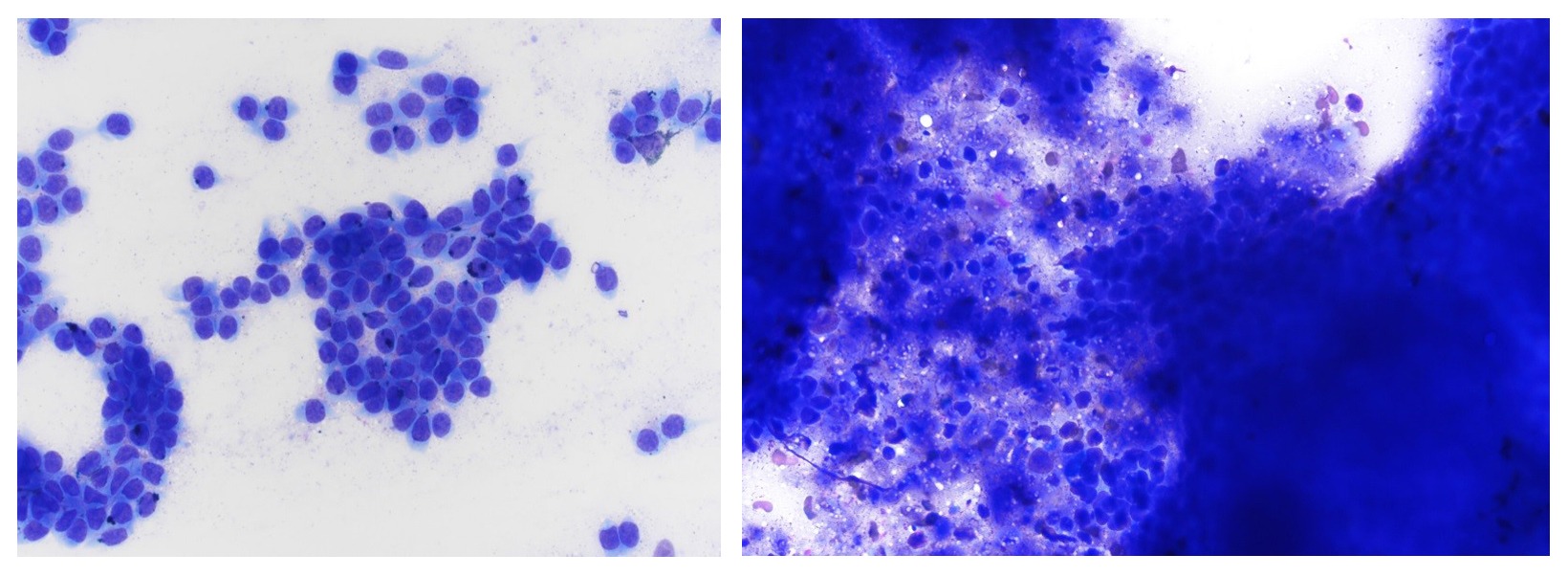
Cutaneous basilar epithelial neoplasm, formerly called basal cell tumor, is now classified histologically by supporting differentiation into epidermis, trichofollicular epithelium, or adnexal structures of apocrine or sebaceous glands.3 Most of the previously diagnosed feline basal cell tumors were likely tumors of hair germ origin such as trichoblastoma or apocrine duct adenomas. Himalayan and Persian cats are at increased risk of developing this type of tumor. They may occur in cats as young as 1 year of age with a peak incidence between 6 and 13 years of age. They occur in the deep dermis and subcutis and are multilobulated, often pigmented cystic tumors that may yield dark brown to black fluid on fine needle aspiration.4 Cytologic appearance is shown in Figure 1.

Figure 1. Aspirates of cutaneous basilar epithelial neoplasms. Left. Note cohesive aggregates of uniform small polygonal cells characterized by a high nuclear-to cytoplasmic ratio, monomorphic round to oval nuclei and a small amount of medium blue cytoplasm with occasional flecks of dark pigment considered to be melanin. Histologic diagnosis: Trichoblastoma (Diff-Quik®, 500x magnification.) Right. Note centrally located amorphous blue cystic material, degenerate cells and scant dark pigment (melanin) surrounded by large blue cohesive aggregates of small epithelial cells. Histologic diagnosis: Apocrine duct adenoma. (Diff-Quik®, 500x magnification.)
Squamous Cell Carcinoma
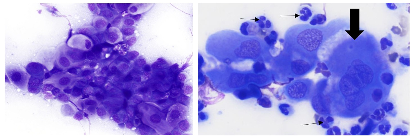
The peak incidence of squamous cell carcinoma in cats is 9-14 years of age.4 Squamous cell carcinoma occurs most commonly in sparsely haired, poorly pigmented skin that has been exposed to UV radiation. Nasal planum, pinnae, and eyelids are the most commonly involved sites. Cats with white facial or pinnal fur have the highest incidence of squamous cell carcinoma.5 Tumors are often locally invasive and may metastasize to regional lymph nodes especially late in the course of disease.3 Imprints of ulcerated masses may yield only septic inflammation, while deep aspirates or scrapings of cleaned and debrided ulcerated areas may yield high numbers of neoplastic epithelial cells. Squamous cell carcinoma can be challenging to differentiate cytologically from hyperplastic or dysplastic squamous epithelium due to inflammation and repair. Histopathology may be required to make a definitive diagnosis, especially of well-differentiated squamous cell carcinoma. If sufficient criteria of malignancy are displayed by the epithelial cells, a presumptive diagnosis of carcinoma can be made via cytologic evaluation. Cytologic appearance is shown in Figure 2.

Figure 2. Aspirates of squamous cell carcinoma. Left. Mass on pinna: Note sheets of adherent oval to angular atypical squamous-type epithelial cells exhibiting an increased nuclear-to-cytoplasmic ratio and moderate anisocytosis and anisokaryosis. (Diff-Quik®, 500x magnification.) Right. Mass on eyelid: Note neutrophilic inflammation (thin black arrows) and individualized dysplastic squamous epithelial cells that exhibit enlarged, irregular and multiple open vesicular nuclei (see arrow), marked anisocytosis and moderate anisokaryosis. (Wright-Giemsa stain, 1000x magnification.)
Mast Cell Tumor
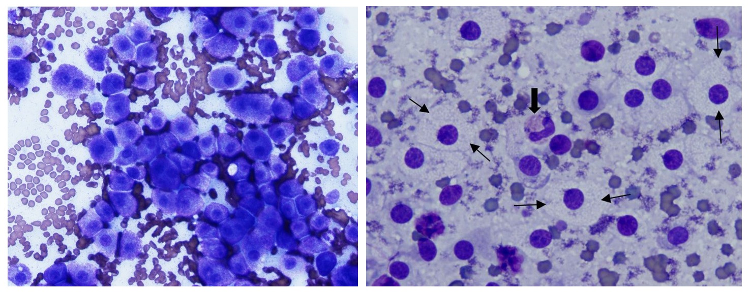
The majority of cutaneous mast cell tumors occur in cats over 4 years of age and the mean age is 10 years.4 Mast cell tumors are a common cutaneous neoplasm of cats that frequently occur as solitary, well-circumscribed masses on the head, neck and limbs. Multiple masses are common in young Siamese cats. The solitary form is generally a benign neoplasm with some exceptions of recurrence and invasion. Features generally associated with more aggressive clinical course in dogs such as nuclear pleomorphism, high mitotic rate and deep dermal invasion have no prognostic significance in cats with solitary tumors. Many young cats with multiple masses respond with spontaneous regression in weeks to months. Aqueous-based stains, such as Diff-Quik®, often do not stain granules well so they may appear as clear vacuoles rather than purple granules in some cases.3 In those cases, consider staining additional slides with methanolic-based Wright’s stain, which may stain the granules.4 Cytologic appearance is shown in Figure 3.

Figure 3. Aspirates of mast cell tumors. Left. Cutaneous mast cell tumor has medium discrete round cells exhibiting mild anisocytosis. Mast cells have 1-2 medium, round central nuclei and numerous small purple granules that fill the abundant cytoplasm. (Diff-Quik®, 500x magnification.) Right. Colonic mast cell tumor showing discrete round cells with non-staining granules that give cells a “fried egg” appearance. Thin black arrows identify cytoplasmic borders of some of the mast cells. Note centrally located round nucleus and abundant cytoplasm with many minute clear vacuoles representing the non-staining granules. Thick black arrow identifies an eosinophil. (Diff-Quik®, 1000x magnification.)
Spindle Cell Sarcoma
Most spindle cell sarcomas in the skin or subcutis of cats are fibrosarcomas, which will be the focus of this review. Fibrosarcomas are typically invasive neoplasms that may adhere firmly to underlying tissue and approximately 25% will metastasize. Vaccine-associated fibrosarcomas, thought to be related to subcutaneous administration of killed vaccines, are frequently high grade and thus very locally invasive and aggressive with a tendency to metastasize.3 They arise at vaccine sites including neck, thorax, lumbar region, flank, and limbs. Since protocols proposed in 1996 recommended right distal limb for feline rabies vaccination, there has been a decrease in interscapular tumors and a significant increase in right hindlimb tumors. This tumor is seen in cats as young as 3 years of age, but the mean age is 8.1 years, which is slightly younger than non-vaccine associated fibrosarcomas (mean 9.2 years).4 Fibrosarcomas may exfoliate cells poorly especially when large amounts of collagen matrix are present, thus incisional biopsy for histopathology may be required for diagnosis. Spindle cell tumors generally cannot be further categorized cytologically and require histopathology and possibly immunohistochemistry for accurate determination of histogenesis. In addition, histologic evaluation may be required to definitively differentiate a reparative or reactive processes such as fibrosis or desmoplasia from a sarcoma. Cytologic appearance is shown in Figure 4.

Figure 4. Cytologic appearance of spindle cell sarcoma. Left. Note medium to large pyriform to spindle-shaped cells occurring singly and in small aggregates. Cells exhibit moderate anisocytosis and anisokaryosis and bizarre mitotic figures (thick black arrow). Thin black arrows identify multinucleated giant cells. Spindle cells have medium, round to oval, slightly eccentric nuclei and tapering blue cytoplasmic processes. Green arrow identifies pink collagenous stroma. Histologic diagnosis: Fibrosarcoma, high grade, presumed vaccine-associated (Diff-Quik®, 600x magnification.) Right. High magnification of neoplastic spindle cells showing coarsely stippled to reticular nuclear chromatin and multiple, small to medium-size, round to irregular dark nucleoli. Histologic diagnosis: Fibrosarcoma, intermediate grade (Diff-Quik®, 1200x magnification.)
Summary
Cutaneous and subcutaneous masses are readily accessible for fine-needle aspiration or deep tissue scrapings and rapid cytologic assessment. The four most commonly reported cutaneous tumors in cats are basilar epithelial tumors, squamous cell carcinoma, mast cell tumor and fibrosarcoma.2 Cytologic examination of cutaneous and subcutaneous tumors, especially epithelial and mast cell tumors, is often rewarding due to the relatively high cell yield on fine-needle aspiration or deep tissue scrapings. Fibrosarcomas may be poorly exfoliating and require incisional biopsy for diagnosis. The results of cytologic evaluation may provide information about prognosis, guidance for next diagnostic steps and/or treatment.
References
- Ghisleni G, Roccabianca P, Ceruti R, et al: Correlation between fine-needle aspiration cytology and histopathology in the evaluation of cutaneous and subcutaneous masses from dogs and cats. Vet Clin Pathol 35(1):24-30, 2006.
- Ho N, Smith KC, Dobromylskyj, MJ: Retrospective study of more than 9000 cutaneous tumors in the UK: 2006-2013. J Feline Med and Surg, vol. 20, 2: pp. 128-134, 2017.
- Raskin RE and Meyer DJ: Canine and Feline Cytology—A Color Atlas and Interpretation, 2nd edition: Saunders Elsevier, Inc. 2010, pp. 46-48, 54-57.
- Meuten D, Editor: Tumors in Domestic Animals, 5th Ames, Iowa: WILEY Blackwell, 2017, pp. 97, 115-116, 145, 195.
- Gross, TL, et al: Skin Diseases of the Dog and Cat—Clinical and histopathological Diagnosis, 2nd Ames, Iowa: Blackwell, 2005, pp. 581-582
- Cowell RL, et al: Diagnostic Cytology and Hematology of the Dog and Cat, 3rd St. Louis, Missouri: Mosby Elsevier, 2008, pp. 100-101.