-
Adopt
-
Veterinary Care
Services
Client Information
- What to Expect – Angell Boston
- Client Rights and Responsibilities
- Payments / Financial Assistance
- Pharmacy
- Client Policies
- Our Doctors
- Grief Support / Counseling
- Directions and Parking
- Helpful “How-to” Pet Care
Online Payments
Emergency: Boston
Emergency: Waltham
Poison Control Hotline
-
Programs & Resources
- Careers
-
Donate Now
 By Brendan Noonan, DVM, DABVP (Avian Practice)
angell.org/avianandexotic
avianexotic@angell.org
617-989-1561
Domestication of O. cuniculus took place between the 5th and 10th centuries, and rabbits have since distributed to every continent save Antarctica. The presence of sharp rostral incisors resulted in their classification alongside rodents. However, their incisor anatomy is also what sets them apart, given that a second pair of tiny upper incisors is present behind the major set. Despite strong similarities between rodents and rabbits, it was determined in the 20th century that the dentition represented an example of divergent evolution that separated rabbits into a separate mammalian order, Lagomorpha. A species that was once raised for meat and fur has since transformed into more than 60 recognized fancy breeds ranging in size, shape and coat. They have since found their way into our homes as pets, but regardless of size or breed, all pet rabbits are susceptible to dental disease. Whether acquired or congenital, their unique anatomy predisposes them to dental disease unlike that seen in dogs and cats.
 Rabbit teeth are classified as elodont (for their continuous growth with no anatomic root) and hypsodont (for having a long crown). The dental formula of the rabbit is 2(I 2/1, C 0/0, PM 3/2, M 3/3) =28. The lack of canine teeth creates an elongated diastema between the incisors and premolars. Rabbit mouths exhibit anisognathism, which means that their lower jaw is narrow when compared to the upper. Rabbits also have a maximum gape of about 20-25 degrees, which makes evaluation of the teeth and dental procedures more difficult. The portion of the tooth that can be visualized above the gum line is called the clinical crown, while the portion that remains below the surface is the reserve crown. Since the teeth are continuously growing, they also do not have a true root, but instead have an apex from which new growth emerges.
The upper incisors consist of hard enamel on the rostral surface and softer dentin along the lingual surface. The differing densities of these surfaces creates a sharp edge as it wears. The lower incisors are covered in enamel on the labial and lingual surfaces, which better suits their normal occlusal placement between the major maxillary incisors and the smaller secondary set of upper incisors, better known as the peg teeth. At rest, the opposing incisors and cheek teeth (premolars and molars) remain in contact. When eating, rabbits will use the incisors to cut the food into manageable lengths using a vertical, chopping motion. Once the food is passed to the cheek teeth, a rotating horizontal stroke is used to grind the food prior to ingestion. This horizontal motion is a large contributor to proper dental wear, which is only achieved when coarse roughage such as hay is consumed. During this process, every lower cheek tooth occludes with 2 upper cheek teeth with the exception of the first mandibular premolar (PM 3) and the last mandibular molar (M 3). This dual occlusion allows for normal wear even in the absence of one opposing cheek tooth.
Any process that impedes the normal eruption and wear of elodont teeth has the potential for causing dental disease, which is divided into four main classes: congenital, traumatic, metabolic bone disease, and abnormal wear. Rabbits born with prognathism or brachygnathism are likely to develop overgrown incisors and potentially abnormal wear of their cheek teeth due to an abnormal pairing of the upper and lower jaw. This is commonly seen in dwarf and lop rabbits. Any traumatic event that alters the jaw or fractures teeth into the alveolar cavity can offset wear or result in infection. Metabolic bone disease is relatively rare, but has been confirmed in several studies where parathyroid hormone was elevated and calcium levels were low. Demineralization of the skull and loosening of the teeth within the alveolar bone causes changes in the occlusal surface while periosteal deformation predisposes to infection.
The most important class of dental disease is acquired through improper wear. Wild rabbits select foods high in fiber and silicates which require exaggerated horizontal movement and appropriate wear of the clinical crown. In captivity, a diet high in grass hay and fresh greens is the best substitute for selections of their wild counterpart but cannot exclude the potential for acquired disease. However, rabbits that lack fiber in their diet are far more likely to end up developing dental disease over time. Without fiber and normal masticatory movements the elodont teeth continue to grow and create pressure on the apex. The apex will be pushed further into the socket, elongating the reserve crown and bending the apex which will change the direction of growth in the clinical crown. Over time lingual points will develop along the mandibular cheek teeth and buccal points will be created on the maxillary cheek teeth as the normal chewing motions cannot wear the exaggerated points. The horizontal chewing motion required to grind hay will aggravate lesions in the mouth and rabbits will begin to prefer pellets that can be crushed using vertical masticatory motions, further exacerbating the problem.

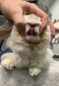
Figure 1. Examining incisors: smooth whiskers back, ensure nares open, part lips, and examine from front and side.
As dental disease begins to affect the patient, owners may notice weight loss, dysphagia, drooling, anorexia, change in fecal size/quantity, excessive salivation, or change in food preferences. While onset of gastrointestinal stasis can have many etiologies, deciding if dental disease is playing a role can typically be determined during the physical exam. Inspection of the incisors can be achieved by gently pulling the upper lips caudally from both sides with special care taken not to occlude the nostrils by pulling the lips dorsally. This will allow you to inspect the occlusion of the incisors from the front and sides, identifying if the lower incisors fit in the notch between the two sets of upper incisors. The limited range of motion of the jaw and narrow gape of the patient makes cheek teeth more difficult to examine. The diastema provides a gap for insertion of appropriate instruments to visualize the cheek teeth. A plastic otoscope cone is a safe tool to use as iatrogenic tooth damage is unlikely if the patient were to chew it. Metal nasal speculums have the benefit of visualizing the lingual and buccal aspects of the teeth simultaneously but should be used by a more experienced handler to avoid a fracture. Subtle abnormalities are likely to be overlooked in a conscious exam but subtle cues like excessive saliva or bubbles can give confidence that a sedated exam should be recommended.
Evaluation of whether the patient can undergo a sedated exam is an important first step. Some animals may benefit from supportive care to ensure they are a better anesthetic candidate. Certain cases also benefit from advanced imaging prior to a sedated dental procedure to help plan the approach. Rabbits with jaw abscesses, exophthalmia, dacryocystitis, or persistent purulent nasal discharge could all have an elongated reserve crown that has dragged bacteria from the mouth into the soft tissues, which sets up infection. Four view skull radiographs are one way to try and elucidate which teeth are contributing to the presenting clinical signs. Computed tomography is the preferred diagnostic modality given the significantly improved information you can gain which helps with treatment planning.
Incisor malocclusion can be treated by either routine trimming of the affected teeth or by extraction. Deciding which plan to offer is based on signalment, history and diagnostic work up. Young rabbits with congenital incisor malocclusion are better candidates for extraction as the frequency and cost of trimming can become overwhelming every 4-6 weeks for the next 7-10 years. Extraction is the only option for rabbits presenting with tooth root infection. Trimming can be performed awake in calm rabbits who allow for proper manual restraint. Sedation or gas anesthesia will be necessary for some rabbits to prevent iatrogenic injury to the surrounding soft tissues during a trim. A DremelTM tool with a diamond wheel is the preferred instrument to reduce the likelihood of longitudinal fractures. A tongue depressor is used on the mesial surface to protect the soft tissues once the disc has transected the incisors. Incisor trimming with nail trimmers, wire cutters or rongeurs should all be used with caution. Crushing the teeth with these tools can lead to longitudinal fractures that will cause introduction of bacteria to the reserve crown and surrounding soft tissues. Incisor extraction is always performed under anesthesia. The use of a Crossley Incisor LuxatorTM as well as flattened and curved large gauge hypodermic needles are required. Ensuring that all the dental pulp is removed is important to reduce the incidence of the incisors growing back in. Using properly curved needles to curettage the alveoli after extraction can reduce the incidence of regrowth.
Molar malocclusion is especially common in patients with reduced hay intake. Molar points can be identified on routine exam in a healthy animal or during examination of an animal with a reduced appetite. Rarely are molar points able to be effectively trimmed in the awake patient. Inhalant anesthesia is required to allow for a patient to be properly placed on a dental board where full examination of the cheek teeth can be performed. A low speed drill is the ideal tool to quickly and effectively reduce molar spurs or malocclusion. Hand tools, such as rongeurs and dental rasp, can be useful in conjunction with a low speed drill but would prolong a dental procedure if used alone. Finding loose or diseased teeth may indicate the need for extraction. Intraoral extraction can prove difficult due to the limited gape of the rabbit mouth (20-25o) and the elongated diastema which places the cheek teeth farther back in the mouth. If indicated, a Crossley Molar ExtractorTM and extraction forceps will be necessary. If only one molar is removed then removal of the opposing tooth is not necessary. Each cheek tooth engages with two opposing teeth and will allow for normal wear.
Dental abscesses are a common sequelae of chronic dental disease. If the teeth are not wearing properly, the pressure from the elongated teeth opposing one another will push the reserve crown toward the apex. This will allow for oral bacteria to be pulled beneath the gum line and create a periapical infection. Dental abscesses can be found along the mandible, inter-mandibular space, cheeks, maxilla, or retrobulbar space. A CT scan is always recommended to determine which teeth are involved so they can be removed during debridement. Without removing these teeth, recurrent abscesses are more likely. Culture and sensitivity of the abscess capsule should be acquired during surgical excision and debridement of the abscess. Marsupialization of the site and flushing daily with dilute povidone iodine allows for healing by second intention. Wound packing with antibiotic soaked umbilical tape or antibiotic impregnated beads has also been described.
Understanding the anatomy and physiology of rabbit dentition can help impress upon clients the important role a diet high in fiber can have. Many of the common dental problems we encounter can be avoided through proper diet alone. Prevention is key since management of improper dental wear requires frequent follow up for the life of the patient.
- Quesenberry, Katherine E., Carpenter, James W. Ferrets, Rabbits, and Rodents Clinical Medicine and Surgery. Louis: Saunders, 2012. Third edition.
- Capello, Vittorio. Rabbit and Rodent Dentistry Handbook. Lake Worth: Zoological Education Network, 2005.
- Bohmer, Estella. Dentistry in Rabbits and Rodents. John Wiley and Sons, 2015
- Gardhouse, S., Sanchez-Migallon Guzman, D., Paul-Murphy, J., Byrne, BA., Hawkins, MG.(2017) Bacterial isolates and antimicrobial susceptibilities from odontogenic abscesses in rabbits: 48 cases. Veterinary Record 181, 538.