-
Adopt
-
Veterinary Care
Services
Client Information
- What to Expect – Angell Boston
- Client Rights and Responsibilities
- Payments / Financial Assistance
- Pharmacy
- Client Policies
- Our Doctors
- Grief Support / Counseling
- Directions and Parking
- Helpful “How-to” Pet Care
Online Payments
Emergency: Boston
Emergency: Waltham
Poison Control Hotline
-
Programs & Resources
- Careers
-
Donate Now
 By Meagan R. Painter, DVM, DACVD
![]()
angell.org/dermatology
dermatology@angell.org
617-524-5733
January 2022
Introduction
Atopic dermatitis is a common, inherited, chronic, relapsing inflammatory and pruritic disease involving skin barrier defects and inflammatory dysfunction. This condition often leads dogs and cats to be presented to the veterinarian for secondary symptoms such as otitis externa, a relapsing bacterial infection of the skin, or unmanageable pruritus.
There are several medical management tools available for veterinarians to treat the secondary symptoms of allergy. Examples include medications like Apoquel (oclacitinib), Atopica (cyclosporine, modified), Cytopoint (lokivetmab), antihistamines, or steroids. However, these therapeutics are designed to mask the symptoms of the disease. They do not influence the source or pathogenesis directly.
Allergen-specific immunotherapy (ASIT) is the practice of administering gradually increasing quantities of allergen extract to an allergic patient with hopes of reducing the symptoms associated with exposure to causative allergens.1
In veterinary medicine, ASIT is a well-established therapy for managing environmentally driven canine and feline atopic dermatitis. Prescriptions for ASIT are unique to a particular patient and based upon results obtained with intradermal allergy testing (IDAT) or in vitro allergy testing (IgE serology).
The pathogenesis of atopic dermatitis involves inflammatory dysfunction and cytokine dysregulation generated after exposure to specific allergenic epitopes. Exposure is percutaneous and results in the binding of allergens to specific IgE receptors located on the surface of antigen-presenting cells. These cells generate an immune response, leading to effector Th2 cells producing various cytokines such as IL-4, IL-5, IL-6, IL-13, and IL-31. Chronic and acute presentations of allergic skin and ear disease will have different inflammatory profiles, which result in various clinical presentations.
However, the hallmark feature of canine and feline atopic dermatitis is the distinct loss of immunologic tolerance. Tolerance is an important feature of the immune system which prevents response against a specific antigen. When tolerance is lost, disorders such as food allergy, auto-immune disease, and atopic dermatitis occur.
This system of tolerance is generally well-orchestrated and straightforward. Antigen-presenting cells, especially dendritic cells, play an essential role in maintaining peripheral tolerance and immunity. Recognition of a foreign protein (antigen) by an antigen-presenting cell leads to cellular migration to a local lymph node. What happens next depends on the environment within the lymph node and the individual’s genetic tendency.
Allergic responses lack tolerance, which results in reduced production of regulatory cytokines and increased production in inflammatory ones.
Allergen-specific Immunotherapy Overview
Allergen-specific immunotherapy (ASIT) has multiple effects on the T-cell-specific response to allergens, and the most important is increasing cytokine production with regulatory activity (IL-10, TGF-b). This can lead to an increase in allergen-specific immunoglobulin (IgG) which can serve as a blocking antibody, outcompeting IgE upon exposure to various allergens.
ASIT remains the only therapeutic for atopic disease, which reshapes the immune response. All other therapeutics restrict inflammatory action by way of blocking or neutralizing cytokines. This critical difference is why ASIT is recommended for most patients with diagnosed canine or feline atopic dermatitis. This therapy treats the source, not just the symptoms.
There are two main types of immunotherapy currently used in veterinary practice.
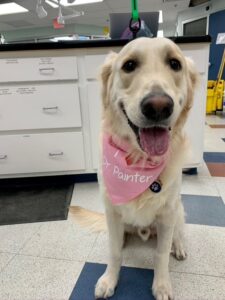
Above is a wonderful Angell-West canine atopic patient who successfully completed ILIT and is now receiving SCIT maintenance.
Subcutaneous immunotherapy (SCIT) has been utilized in dogs and cats for many decades. This involves injecting an aqueous, saline-phenol preserved extract in two or three-vial sets of increasing concentration until a maintenance injection dose and frequency is reached.
Sublingual immunotherapy (SLIT) is an oral modality that harnesses oromucosal dendritic cell potential to induce tolerance. A measured drop of glycerinated antigen extract is administered into the oral cavity (cheek pouch) once or twice daily. Given their relative abundance in the oral mucosa, and their default nonreactivity to substances placed in the mouth (e.g food), this delivery method is promising for both dogs and cats.
There are distinct advantages and disadvantages to each type of ASIT in veterinary medicine. Formal clinical trials are difficult to perform. As a result, published comparisons of clinical outcomes with SCIT and SLIT are lacking in the veterinary arena.
Intralymphatic Immunotherapy Overview
Intralymphatic immunotherapy (ILIT) is an evolving form of allergen-specific immunotherapy involving the injection of allergen extract directly into the lymph node.
This process introduces lower doses of allergen to many highly immunocompetent lymphocytes located with a specific lymph node. The popliteal lymph node is selected and identified in dogs and cats through manual or ultrasound identification. As a result of this direct exposure, the probability for tolerance induction is maximized. Cumulative doses are about 1,000 times lower than SCIT doses. Adverse event risk is, therefore, lower.
After patients complete the ILIT induction protocol, they are transitioned to SCIT or SLIT for maintenance.
It remains to be seen if this type of immunotherapy delivery is more efficacious when compared to SCIT and SLIT alone. More extensive studies in a clinical context are needed. At this time, only a handful of veterinary dermatology clinics in the United States (including Angell-West, Waltham, MA) are utilizing intralymphatic immunotherapy in both canine and feline atopic patients.
The potential for this desensitization method to be clinically useful, safe, and efficacious for our patients is exciting and pioneering.
Goals and Next Steps
The goal with ASIT is to reduce medication dependence or symptom severity for patients suffering from canine or feline atopic dermatitis. It is essential to recognize that improvement in patients is individualized and often on a spectrum. Some patients have excellent control of their atopic disease and can come off anti-pruritic therapeutics entirely. Other patients have moderate control of their atopic disease and can lessen their medication needs. And, yet, others fail to respond to ASIT entirely.
Given the positive safety margin of ASIT and the potential to produce profound immunologic effects on the atopic individual, this therapeutic modality should be considered for most atopic patients.
It is imperative to approach every patient with atopic disease from a multimodal perspective. For example, medical management options provide relief from symptoms and improve life quality. Results are often fast and reliable. We are fortunate to practice in a time when targeted therapeutics are available for our patients with atopic disease.
The value of ASIT from a clinical perspective is potentially massive. If a patient can reduce their medication dependence or symptom severity because of a specific desensitization protocol, the gold standard goal for managing the atopic disease has been reached.
However, ASIT can indeed take weeks to months to usher clinical improvement. Similarly, schedules and prescriptions are individualized and based on a myriad of factors taken into consideration by veterinary dermatologists with advanced training in ASIT prescribing and maintenance practices.
Referral to a board-certified veterinary dermatologist for allergen-specific immunotherapy remains the standard of care for canine and feline atopy. This ensures the patient is diagnosed correctly, medically managed, and started on a targeted ASIT protocol designed specifically for them.
Yet, there is no doubt that management of canine and feline atopic disease is done best when primary and specialty veterinarians work together to advance clinical outcomes and maintain patient life quality and safety. This is undoubtedly an exciting, collaborative time in veterinary practice – and these advances in immunotherapy showcase some of the value of advanced clinical care for this common disease.
References
- The future of immunotherapy for canine atopic dermatitis: a review. DeBoer, Douglas. Veterinary Dermatology, 2017, 28: 25-e6.
- Update on the pathogenesis, diagnosis, and treatment of atopic dermatitis in dogs. Nuttall, Tim et al. JAVMA, 2019. Vol 254, 1291-1300.
- A review of allergen-specific immunotherapy in human and veterinary medicine. Mueller, Ralf et al. Veterinary Dermatology, 2009. 20(2): 84-98.
- Allergen immunotherapy in people, dogs, cats, and horses – differences, similarities, and research needs. DeBoer, Doug et al. allergy. 2018. Oct; 73(10): 1989-1999.