-
Adopt
-
Veterinary Care
Services
Client Information
- What to Expect – Angell Boston
- Client Rights and Responsibilities
- Payments / Financial Assistance
- Pharmacy
- Client Policies
- Our Doctors
- Grief Support / Counseling
- Directions and Parking
- Helpful “How-to” Pet Care
Online Payments
Referrals
- Referral Forms/Contact
- Direct Connect
- Referring Veterinarian Portal
- Clinical Articles
- Partners in Care Newsletter
CE, Internships & Alumni Info
CE Seminar Schedule
Emergency: Boston
Emergency: Waltham
Poison Control Hotline
-
Programs & Resources
- Careers
-
Donate Now
 By Jennifer Michaels, DVM, DACVIM (Neurology)
![]()
angell.org/neurology
neurology@angell.org
617-541-5140
March 2023
x
x
 By Jillian Walz, DVM, DACVIM (Medical Oncology), DACVR (Radiation Oncology)
angell.org/oncology
oncology@angell.org
617-541-5136
x
x
x
Although brain tumors are the most common cause of new-onset neurological symptoms in older dogs, it is important to keep an open mind about differential diagnoses in these patients. Some people, pet owners, and veterinarians alike will assume that simply because a dog is senior, any new onset neurological symptoms must be due to a brain tumor. Subsequently, owners may believe that their pet has few or no treatment options. This article aims to shed light on when an intracranial neoplasia should be suspected versus other causes for new onset neurological symptoms and showcase the myriad treatment options available for presumptive or definitive diagnosis of intracranial neoplasia.
Clinical Signs and Incidence of Brain Tumors
The most common clinical signs of brain tumors depend on location. For supratentorial tumors (cerebral hemispheres, thalamus), seizures and behavioral changes are the most common clinical signs (Foster et al., 1988; Rossmeisl et al., 2013). In one report, in 75% of dogs diagnosed with a supratentorial brain tumor, a seizure was the first reported clinical signs (Schwartz et al., 2011). Central vestibular dysfunction is the most common clinical sign of infratentorial tumors (brainstem and cerebellum) (Rossmeisl et al., 2013). Most dogs with brain tumors have abnormal neurological examinations on presentation. However, up to 64% of dogs with new-onset seizures that were ultimately diagnosed with a structural brain lesion (such as a tumor) had an otherwise normal neurological examination (Pákozdy et al., 2008).
The clinical course of dogs with brain tumors is invariably progressive. Typically, the progression is subacute (weeks) to chronic (weeks to months). For example, the average time between developing new seizures and/or behavioral changes and detecting a persistently abnormal neurological exam is reported to be 78 days (range two to 400 days) (Foster et al., 1988; Rossmeisl et al., 2013).
Differential Diagnoses
There are many differential diagnoses other than brain tumors for new onset and/or progressive, centrally localizing neurological signs in an older dog. Some of the most important are described here with some clinical features that may help prioritize them on your differential list.
Epilepsy of Unknown Cause: Similar to Idiopathic Epilepsy, these patients have recurrent seizures without any other neurological symptoms or deficits; however, they fall outside of the IE diagnostic criteria due to the age of seizure onset. Neurological examination is normal. This accounts for about 1/3 of dogs greater than six years of age presenting with seizures as the chief complaint (Hall et al., 2020)
Canine Cognitive Dysfunction Syndrome: CCDS is a geriatric onset, chronic, slowly progressive neurodegenerative disease akin to dementia in people. Signs typically start with fairly mild behavioral changes such as increased anxiety, pacing, or restlessness at night and progress over months to years. Seizures and asymmetric symptoms such as circling are possible sequelae of CCDS.
Ischemic stroke: Strokes can occur anywhere in the brain and thus cause a variety of neurological signs, although the most common is vestibular syndrome. Clinical signs are per-acute onset, non-progressive, and usually spontaneously improve with time (typically hours to days for initial improvement). Strokes can recur, especially in dogs with underlying risk factors such as hypertension, hypothyroidism, renal disease, or Cushing’s disease.
Diagnosis
Definitive diagnosis of intracranial neoplasia does require advanced imaging, typically brain MRI. While tissue biopsy is required for a definitive diagnosis, 90% or more of cases were correctly diagnosed with an intracranial neoplasm based on MRI (Ródenas et al., 2011).
In many instances, pet owners cannot or do not want to pursue advanced imaging or a definitive diagnosis for the cause of their pet’s neurological symptoms. In these cases, clinical factors that can strengthen the presumptive diagnosis of a brain tumor, including:
- Geriatric patient
- New onset, acute to chronic symptoms (typically more subacute to chronic)
- Progression of symptoms over days to months
- Lack of improvement over time
- Definitive central nervous system signs
- Lateralizing, focal neurological symptoms
- Clinical improvement with empirical steroid therapy (see below)
Palliative Care
 Palliative care for a brain tumor plays a significant role in the management of both definitively diagnosed and presumptive brain tumors.
Anti-inflammatory steroids are the mainstay of palliative therapy. While steroids will generally not treat the tumor directly (excepting round cell neoplasia such as CNS lymphoma), they will reduce peri-lesional edema in the brain, significantly reducing clinical signs and improving quality of life. The recommended starting dosage is 1mg/kg/day of prednisone with a gradual taper over a few weeks. Most brain tumor patients will not tolerate being tapered completely off steroids without a recurrence of their clinical signs. The goal is to find the lowest effective dosage that maintains a good clinical state and minimizes clinical deficits. Given that this therapy is not treating the tumor, clinical signs will inevitably return as the tumor grows. When deficits progress, the prednisone dose can be incrementally increased until it ceases to control the deficits or the patient experiences dose-limiting side effects.
Even for owners who are unable or do not wish to pursue a definitive diagnosis for the cause of their pet’s neurological signs, if the index of suspicion is high, it is very reasonable to consider at least a trial of anti-inflammatory steroids. Although there is no guarantee that the patient will improve, most dogs with a brain tumor will show some improvement with steroids, which should raise the index of suspicion for a presumptive brain tumor diagnosis and support continued steroid treatment.
Palliative care also importantly includes symptomatic therapy and supportive care, as needed. This includes maintenance anti-seizure therapy for patients experiencing seizures. While any maintenance anti-seizure medication can be utilized, this author prefers levetiracetam (20-30 mg/kg PO q8h), levetiracetam ER (30 to 45 mg/kg PO q12h), or zonisamide (5 to 10 mg/kg PO q12h) over the older generation anti-seizure medications such as phenobarbital or potassium bromide due to preferable side effect profile. Other important supportive care therapies could include anti-nausea medications such as maropitant (6 to 8 mg/kg PO q24h) or ondansetron (0.5 to 1 mg/kg PO q8-12h) for patients with vestibular signs or appetite stimulants, as needed.
As with any of the treatments discussed in this article, prognosis with palliative care depends somewhat on tumor type and location. However, in general, the median survival time for palliatively treated brain tumors is two to three months, with forebrain tumors being associated with a longer survival time (median: five to six months) compared to brainstem tumors (median: one month) (Hu 2015, Rossmiesl 2013).
Surgery
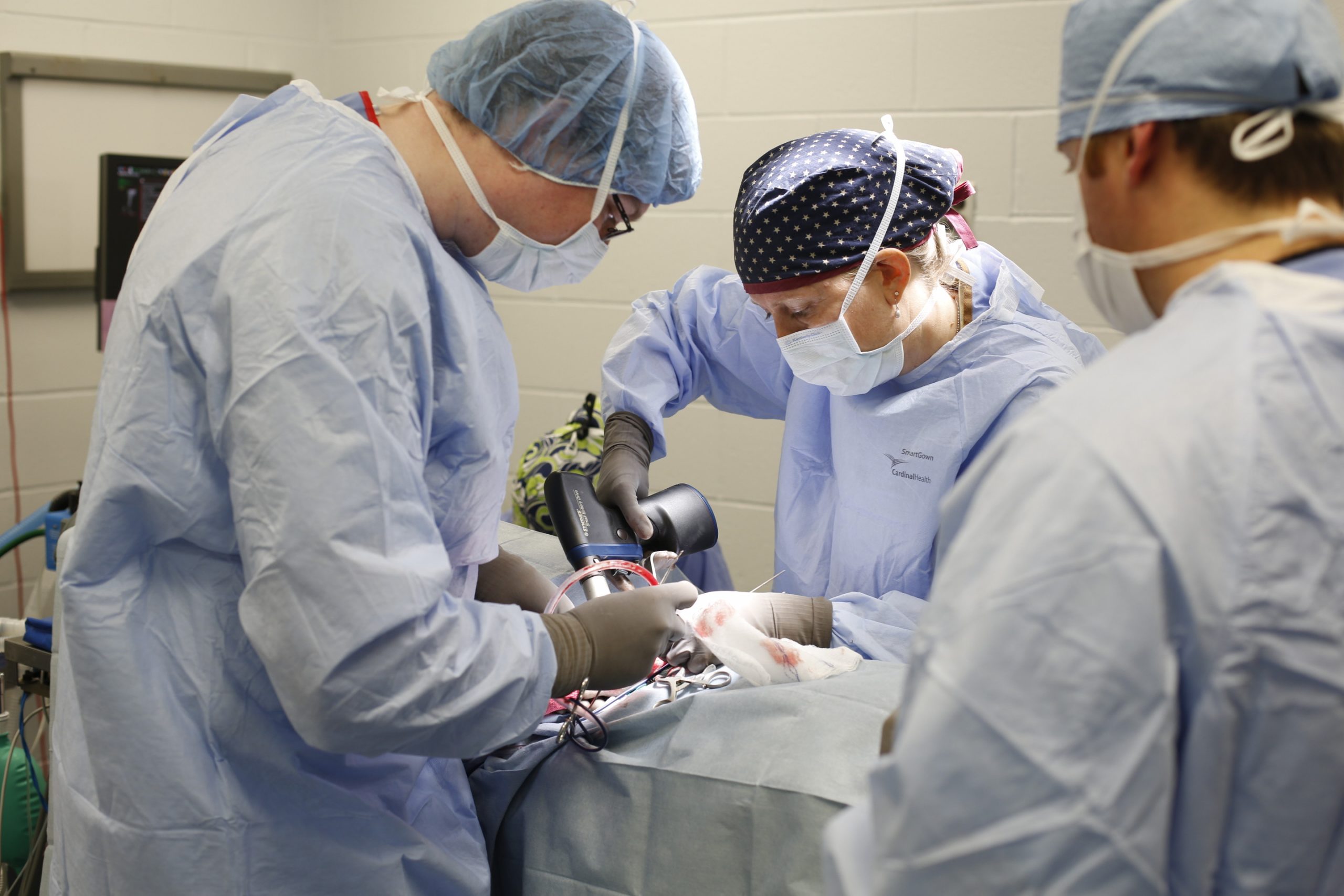
The benefit of surgical treatment includes rapid decompression for those patients with large masses or masses causing marked mass effects with concern for herniation and acute decompensation as well as histologic confirmation of tumor type, thus potentially allowing for more targeted therapy options. However, given the inherent degree of invasiveness, surgery could easily be considered the riskiest of treatment options. Surgery has a mortality rate of up to 13%, and a major complication rate (e.g., worsening of neurological status, new or worsening seizures, aspiration pneumonia) of up to 18% has been reported (Kohler et al. 2018).
Surgery may not be a viable treatment option for all dogs with a brain tumor. The primary limiting factor to the utility of surgery is tumor location. In general, infratentorial and intra-axial tumors are less likely to be surgical candidates than rostral fossa or extra-axial tumors. Surgical approaches for more difficult-to-access tumors, including those of the brainstem, pituitary, and peri-ventricular regions, have been described, although they are less commonly performed and often require specialized equipment and training not available at all institutions.
MST for surgical treatment of primary brain tumors is 10.5 months (one to 70 months) (Hu et al. 2015). Meningioma, the most common primary brain tumor in dogs, has a median survival time of approximately nine months when treated with surgery alone.
While there is clearly some benefit to considering surgery, it is less often pursued as an isolated treatment option given the availability, reduced invasiveness, and improved outcomes associated with radiation therapy (see below). However, there are certain patients or circumstances in which surgery can be beneficial, especially if followed up with adjuvant oncologic therapy.
x
References
- Hu H, Barker A, Harcourt-Brown T, Jeffery N. Systematic Review of Brain Tumor Treatment in Dogs. J Vet Intern Med. 2015;29(6):1456-1463. doi:10.1111/jvim.13617
- Rossmeisl JH Jr, Jones JC, Zimmerman KL, Robertson JL. Survival time following hospital discharge in dogs with palliatively treated primary brain tumors. J Am Vet Med Assoc. 2013;242(2):193-198. doi:10.2460/javma.242.2.193
- Hall R, Labruyere J, Volk H, Cardy TJ. Estimation of the prevalence of idiopathic epilepsy and structural epilepsy in a general population of 900 dogs undergoing MRI for epileptic seizures. Vet Rec. 2020;187(10):e89. doi:10.1136/vr.105647
- Foster ES, Carrillo JM, Patnaik AK. Clinical signs of tumors affecting the rostral cerebrum in 43 dogs. J Vet Intern Med. 1988;2(2):71-74. doi:10.1111/j.1939-1676.1988.tb02796.x
- Kohler RJ, Arnold SA, Eck DJ, Thomson CB, Hunt MA, Pluhar GE. Incidence of and risk factors for major complications or death in dogs undergoing cytoreductive surgery for treatment of suspected primary intracranial masses. J Am Vet Med Assoc. 2018;253(12):1594-1603. doi:10.2460/javma.253.12.1594
- Pákozdy A, Leschnik M, Tichy AG, Thalhammer JG. Retrospective clinical comparison of idiopathic versus symptomatic epilepsy in 240 dogs with seizures. Acta Vet Hung. 2008;56(4):471-483. doi:10.1556/AVet.56.2008.4.5
- Ródenas S, Pumarola M, Gaitero L, Zamora A, Añor S. Magnetic resonance imaging findings in 40 dogs with histologically confirmed intracranial tumours. Vet J. 2011;187(1):85-91. doi:10.1016/j.tvjl.2009.10.011
- Rossmeisl JH Jr, Jones JC, Zimmerman KL, Robertson JL. Survival time following hospital discharge in dogs with palliatively treated primary brain tumors. J Am Vet Med Assoc. 2013;242(2):193-198. doi:10.2460/javma.242.2.193
- Schwartz M, Lamb CR, Brodbelt DC, Volk HA. Canine intracranial neoplasia: clinical risk factors for development of epileptic seizures. J Small Anim Pract. 2011;52(12):632-637. doi:10.1111/j.1748-5827.2011.01131.x