-
Adopt
-
Veterinary Care
Services
Client Information
- What to Expect – Angell Boston
- Client Rights and Responsibilities
- Payments / Financial Assistance
- Pharmacy
- Client Policies
- Our Doctors
- Grief Support / Counseling
- Directions and Parking
- Helpful “How-to” Pet Care
Online Payments
Emergency: Boston
Emergency: Waltham
Poison Control Hotline
-
Programs & Resources
- Careers
-
Donate Now
 By Nicolas J. Trout, MA, VET, MB, DACVS, ECVS
![]()
angell.org/surgery
surgery@angell.org
617-541-5048
January 2022
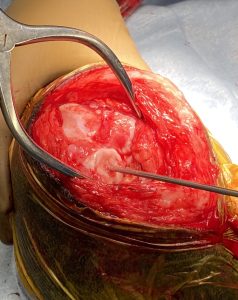
Figure 1
Osteochondrosis (OC) is caused by a failure of normal endochondral ossification leading to abnormal cartilage development. In some cases, this diseased cartilage will separate from the underlying bone to create a flap (Osteochondritis dissecans or OCD). Regardless, these types of articular defects, found in the canine shoulder, elbow, hip, stifle, and hock, frequently occur in growing large breed dogs, leading to lameness, joint pain, joint swelling, and decreased range of motion in the affected joint. Most dogs present between six and twelve months of age, but OC lesions can become a clinical problem later in life. Diagnosis is often straightforward through radiography or computed tomography; however, treatment can be challenging. Historically, the cartilage defect would be curetted down to healthy bleeding subchondral bone, allowing the formation of fibrocartilage. Unfortunately, in the stifle, where the problem lies on the weight- bearing surface of the femoral condyle, positive outcomes are difficult to achieve. The use of a synthetic cartilage plug—SynACART® (Arthrex)—offers an appealing alternative. The plug is made from a biodurable polycarbonate urethane on the articular surface and molded onto a titanium mesh at the base (Figure 1, right), allowing for bone ingrowth and fusion. Plugs come in various sizes: 8mm, 10mm, and 15mm (size of defect can be measured from radiographs or CT scan), which avoids the need for sampling at a donor site, thereby reducing morbidity and speeding up the surgical process.
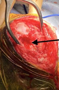
Figure 2
Routine stifle arthrotomy, judicious retraction, and hyperflexion of the stifle allow exposure of the lesion (Figure 2, left). Using a specific guide, a 2.4mm guide pin is placed in the center of, and perpendicular to, the defect (retaining a rim of normal hyaline cartilage). A cannulated drill is placed over the pin and advanced to a stop limited depth of 8mm (Figure 3, below).
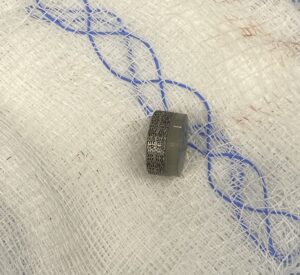
Figure 3
This is carefully flushed and debrided before implantation of the SynACART® plug (Figure 4, below). Postoperative analgesia is the same as for any other stifle arthrotomy with the use of nonsteroidal anti-inflammatories for two weeks. Confinement to a single room is recommended for the first two to four weeks, with five-minute leash walks starting at four weeks. A six-week recheck with radiographs or CT scan is recommended, followed by an incremental return to normal activity at around 12 weeks post-surgery.
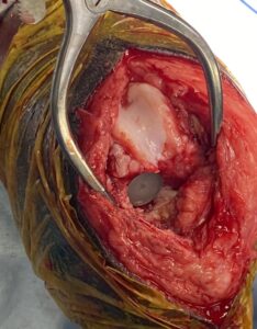
Figure 4
Based on several peer-reviewed scientific articles, it appears as though synthetic osteochondral resurfacing techniques offer a viable alternative to curettage with good to excellent clinical outcomes.
References
- Canine Elbow Dysplasia: Ununited Anconeal Process, Osteochondritis Dissecans, and Medical Coronoid Process Disease. Vezzoni A, Benjamino K. Vet Clin North Am Small Anim Pract. 2021 Mar;51 (2) 439-474.
- Treatment of Osteochondrosis Dissecans of the Canine Stifle Using Synthetic Osteochondral Resurfacing. Egan P, et al. Vet Comp Orthop Traumatol. 2018 Feb;31(2): 144-152.
- Synthetic osteochondral resurfacing for treatment of large caudocentral osteochondritis dissecans lesions of the humeral head in 24 dogs. Murphy S, et al. Vet Surg 2019 Jul; 48 (5): 858-868.
- Evaluation of synthetic osteochondral implants. Cook JL, et al. J Knee Surg. 2014 Aug:27 (4): 295-302.