-
Adopt
-
Veterinary Care
Services
Client Information
- What to Expect – Angell Boston
- Client Rights and Responsibilities
- Payments / Financial Assistance
- Pharmacy
- Client Policies
- Our Doctors
- Grief Support / Counseling
- Directions and Parking
- Helpful “How-to” Pet Care
Online Payments
Emergency: Boston
Emergency: Waltham
Poison Control Hotline
-
Programs & Resources
- Careers
-
Donate Now
 By Nick Trout, MA, VET MB, DACVS, ECVS
angell.org/surgery
617-541-5048
Osteochondrosis (OC), a developmental disorder, is characterized by a failure of normal endochondral ossification of epiphyseal cartilage, seen in young, rapidly growing dogs. Though we tend to focus on the more common cartilaginous defects of the elbow, shoulder, and hock, on occasion the unforgiving stifle joint can be affected. When young, large, or giant breed dogs present with a progressive hind limb lameness, stifle joint swelling and pain on stifle manipulation, be sure to consider stifle OC, a differential ideally diagnosed with plain radiographic changes on the weight-bearing surface of the femoral condyle (Fig. 1a and 1b). Defects can occur on either the lateral or medial femoral condyles with lateral reported to be more common, and it is always worthwhile radiographing the contralateral limb to assess for bilateral disease.
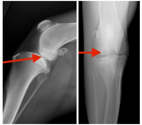
Figures 1a and 1B: Lateral and anterior/posterior plain radiograph of stifle, one-year-old spayed female deerhound. Red arrow defines the medial condylar OC lesion in both views.
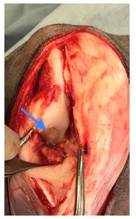
Figure 2: Following lateral stifle arthrotomy, visualization (blue arrow) of the OC lesion.
Unfortunately, treatment options for stifle OC can be extremely frustrating. Conservativemanagement consists of exercise restriction, anti-inflammatories, use of external support and physical therapy, but comes with an anticipation of progressive osteoarthritis and decreased function. Similarly, surgical curretage of the lesion, via arthrotomy or arthroscopy, carries a guarded to poor prognosis, thanks to loss of articular cartilage and damage to subchondral bone, over a relatively large, high load surface of the stifle joint.
Osteochondral autografts have been used in the treatment of canine OC for many years, the stifleoffering excellent locations to harvest healthy articular cartilage away from the weight-bearing surface. The OATS system (Osteochondral Autograft Transfer System, Arthrex Inc, Naples, FL) provides an inexpensive and simple way to prepare the OC defect, obtain appropriately sized graft and complete transplantation.
In the case of an eighteen month old, spayed female deerhound, a lateral stifle arthrotomy was performed, defining the medial condylar OC lesion (figure 2). The defect was measured using a template as 10mm diameter (6, 8 and 10mm OATS are available). Using a donor harvester trephine articular cartilage and subchondral bone graft was obtained (figures 3 and 4). A core of 8-15mm depth is considered ideal (figure 5). The OC lesion/recipient bed is prepared to accommodate the core’s depth (Figures and 6 and 7) and the graft transfer completed (fig 8. 2120).
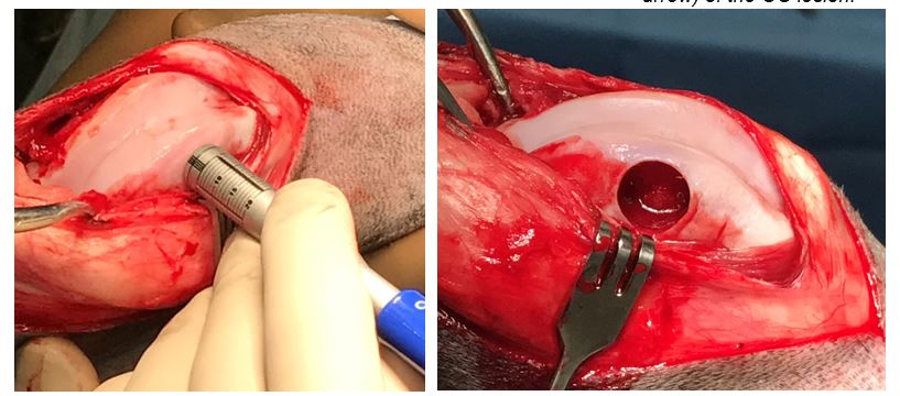
Figure 3 (left) Preparation of the donor graft from non-weight bearing articular cartilage of the lateral trochlear groove. Figure 4 (right) Defect in the trochlear groove will heal by second intention, filling in with fibrocartilage.
Use of OATS in the treatment of stifle OC can result in decreased lameness and improved quality of life but the results are far from perfect. Residual exercise induced lameness and progression of osteoarthritis persists in many cases despite this attempt to restore cartilage and surface integrity. Complications of patella luxation and cruciate injury have been reported. However, until alternative options are available, OATS offers an inexpensive (each OATS kit costs about $150 and can be resterilized), straight forward and positive treatment option in select cases, for a frustrating developmental disorder in growing dogs.
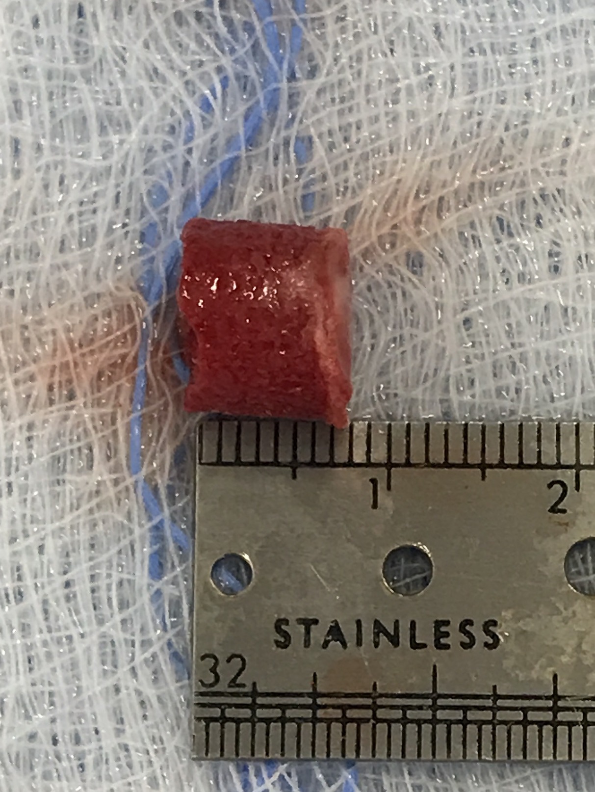
Figure 5: Approximately 8mm X 8mm osteochondral autograft.
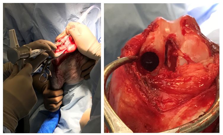
Figure 6 (left): Resection of the OC lesion and preparation of the recipient bed. Figure 7 (right): Recipient bed prepared to accept graft.
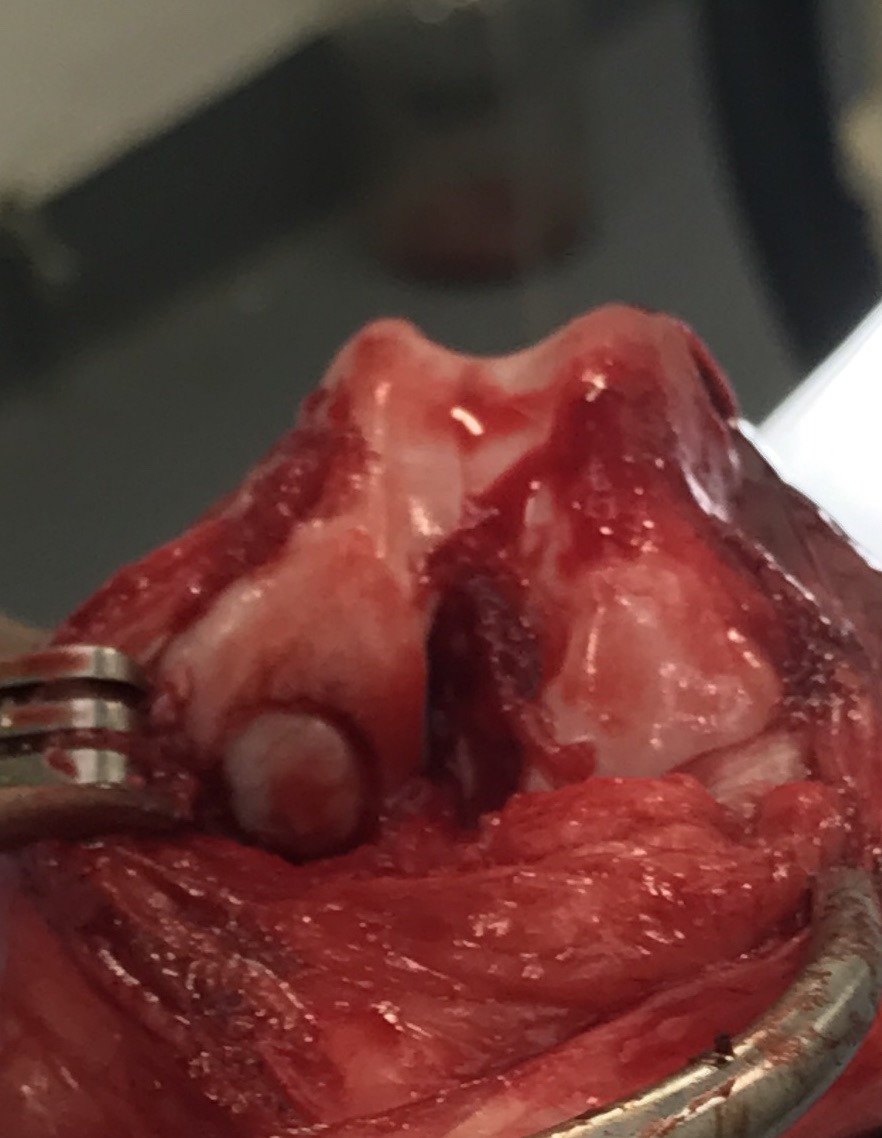
Fig. 8 Osteochondral graft complete. The cartilage surface should be slightly ‘proud’ to avoid ‘counter-sinking’.
References:
Autogenous Osteochondral Grafting for Treatment of Stifle Osteochondrosis in Dogs. Cook JL, Hudson CC, Keiichi Kuroki. Veterinary Surgery, 37:311-321, 2008.
Osteochondral autograft transfer for the treatment of osteochondritis dissecans of the medial femoral condyle in dogs. Fitzpatrick N, Yeadon R, van Terheijden C, Smith TJ. Vet Comp Orthop Traumatol, 25: 135-143, 2012.