-
Adopt
-
Veterinary Care
Services
Client Information
- What to Expect – Angell Boston
- Client Rights and Responsibilities
- Payments / Financial Assistance
- Pharmacy
- Client Policies
- Our Doctors
- Grief Support / Counseling
- Directions and Parking
- Helpful “How-to” Pet Care
Online Payments
Referrals
- Referral Forms/Contact
- Direct Connect
- Referring Veterinarian Portal
- Clinical Articles
- Partners in Care Newsletter
CE, Internships & Alumni Info
CE Seminar Schedule
Emergency: Boston
Emergency: Waltham
Poison Control Hotline
-
Programs & Resources
- Careers
-
Donate Now
 Michele James, DVM, DACVIM (Neurology)
neurology@angell.org
angell.org/neurology
617-541-5140
Overview
Otitis media, an inflammatory disease process affecting the middle ear, is a frequent ailment recognized in small animal veterinary practices in both canine and feline patients. It results in damage to the tympanic membrane and bulla and a variety of clinical signs ranging from pain to vestibular signs. Otitis media can stem from either primary or secondary factors. In canine patients, otitis media typically results secondarily from chronic otitis externa that extends through the tympanic membrane. However, it can also result from primary factors, such as a foreign body, primary secretory otitis media (PSOM) of Cavalier King Charles Spaniels, or iatrogenic rupture during ear cleaning. In contrast, otitis media in feline patients commonly results from primary factors, namely ascending viral, Mycoplasma, or Bordetella respiratory infections via the eustachian tube. Secondary factors for feline otitis media include nasopharyngeal polyps, chronic ear mite infestations, or chronic otitis externa. Untreated or improperly treated otitis media can lead to otitis interna, an inflammatory disease process affecting the inner ear structures, which results in damage to the hearing apparatus and oftentimes deafness and neurologic signs. In severe cases of otitis interna, life-threatening otogenic intracranial infections and meningitis can result via passage of infectious organisms from the inner ear to the brainstem through the nerves and vessels located in the internal acoustic meatus.
Presentation
Patients with otitis media can have variable clinical signs, though many patients may present with complaints of head shaking and aural exudate. During physical examination, mucoid discharge in the external ear canal may be noted. It is important to note that mucus is not produced along the external ear canal, but is produced by goblet cells in the middle ear and eustachian tube. Therefore, the presence of mucus in the external ear canal is consistent with a tear or rupture of the tympanic membrane. Due to local inflammation and swelling around the bulla and nearby temporomandibular joint, secondary to otitis media, pain on palpation of the base of the ear and on opening the mouth, respectively, may occur. This local inflammation may also result in a number of neurologic deficits due to inflammation of the nerves that course through and near the bulla. Keratoconjunctivitis Sicca (KCS) may result from damage to the parasympathetic branch of the facial nerve that innervates the lacrimal gland. Damage to the sympathetic nerves that course through the middle ear may result in ipsilateral Horner’s Syndrome (i.e. miosis, enophthalmos, and ptosis). Damage to the facial nerve may result in ipsilateral facial paresis/paralysis, namely facial asymmetry and inability to blink. Otitis interna may result in damage to the vestibulocochlear nerve and secondary hearing deficits and/or vestibular signs, including a head tilt, nystagmus, and ataxia. A positional ventral strabismus may also be noted, such that when the patient’s head is raised, an eye drop will be noted ipsilateral to the lesion. In severe cases of otitis interna that have progressed to an intracranial infection, mentation change, neck pain, ataxia, paresis, proprioceptive deficits, and seizure activity may be present.
Diagnosis
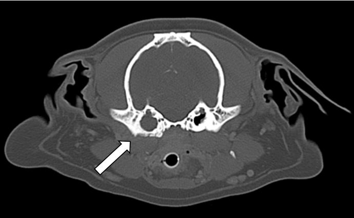
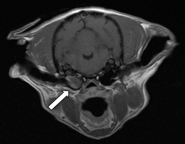
Otitis media can be confirmed through otoscopic examination by visualizing a ruptured or discolored tympanic membrane or mucoid discharge in the ear canal. If there is marked inflammation or stenosis of the ear canal that prohibits otoscopic examination, some practitioners may elect to treat with a 1-2 week course of topical or systemic anti-inflammatory steroids. With the introduction of video otoscopes, otoscopic examination has become much easier and may facilitate otoscopic examination even in a stenotic ear. For instance, one way to evaluate the integrity of the tympanic membrane outside of direct visualization is to fill the ear canal with saline while the patient is in lateral recumbency and watch for air bubbles to rise as the patient breathes. If this is noted, then a tear in the tympanic membrane is present and is allowing air to pass from the nasopharynx through the eustachian tube into the tympanic bulla and through the middle ear. Otitis media/interna may also be diagnosed via imaging, including radiography to detect osseous changes to the bulla. CT (Figure 1) and MRI (Figure 2) are more sensitive diagnostic testing options and can be particularly useful in patients exhibiting neurologic signs. It should be stated that in 70% of otitis media cases the tympanic membrane may be intact. Therefore, diagnosis may also be established on clinical suspicion based on a patient’s history and exam findings, as well as via cytology and culture results obtained from sampling of the middle ear via myringotomy. In patients where otitis interna is suspected, cerebrospinal fluid analysis under general anesthesia may also be beneficial to further evaluate and diagnose meningitis following MRI.

Figure 1. Post-contrast axial view of the skull at the level of the bullae from the head CT of an 11 year old female spayed pug with right otitis media. The right tympanic bulla is filled with isoattenuating material.
Treatment and Prognosis
Antibiotic therapy for otitis media/interna should be determined by the cytology and bacterial culture and susceptibility results from submitted sampling of the middle ear. It is recommended that both aerobic and anaerobic bacterial cultures be performed, as anaerobic infections can occur in up to 25% of patients with a history of chronic otitis. Cytology is recommended to screen for yeast organisms that fail to grow on culture. Steroids or non-steroidal anti-inflammatories may also be prescribed, in conjunction with targeted antibiotic therapy, in order to reduce inflammation and pain associated with otitis media/interna. Antibiotic therapy for otitis media cases should be continued for 6-8 weeks, and in cases where otitis interna or intracranial involvement is suspected antibiotic therapy should be carried out for at least 3 months. Regular rechecks are recommended throughout treatment to ensure an appropriate response to therapy is being achieved.
A majority of otitis media/interna cases can be successfully treated with medical management. Though complete resolution of the infection can typically be achieved, it should be noted that secondary facial paresis/paralysis and head tilt may be permanent deficits, especially if still present 3-4 weeks after starting treatment. In recurrent cases of otitis media/interna or in cases refractory to medical management, surgical intervention with a total ear canal ablation and bulla osteotomy should be pursued.

Figure 2. T1-weighted post-contrast axial view at the level of the caudal colliculus from the brain MRI of a 13 year old female spayed Shih Tzu with right otitis media. The right tympanic bulla is filled with T1 weighted isointense material with heterogeneous mild contrast enhancement (arrow).
References
- Bloom P. Diagnosis and management of otitis media. DVM360.com. April 01, 2009.
- Gotthelf LN. Diagnosis and treatment of otitis media in dogs and cats. Veterinary Clinics Small Animal Practice. 2003; 34:469-487.
- Kennis RA. Feline otitis: diagnosis and treatment. Veterinary Clinics Small Animal Practice. 2013; 43: 51-56.
- Moriello KA. Overview of otitis media and interna. Merck Veterinary Manual. 2018.
- Nanai B, Lyman R. Otitis media and interna: look for neurologic signs. DVM360.com. August 01, 2016.