-
Adopt
-
Veterinary Care
Services
Client Information
- What to Expect – Angell Boston
- Client Rights and Responsibilities
- Payments / Financial Assistance
- Pharmacy
- Client Policies
- Our Doctors
- Grief Support / Counseling
- Directions and Parking
- Helpful “How-to” Pet Care
Online Payments
Referrals
- Referral Forms/Contact
- Direct Connect
- Referring Veterinarian Portal
- Clinical Articles
- Partners in Care Newsletter
CE, Internships & Alumni Info
CE Seminar Schedule
Emergency: Boston
Emergency: Waltham
Poison Control Hotline
-
Programs & Resources
- Careers
-
Donate Now
 By Steven Tsai, DVM, DACVR
angell.org/diagnosticimaging
diagnosticimaging@angell.org
617-541-5139
Acute vomiting is one of the most common reasons for bringing a pet to the veterinarian, and there are innumerable potential causes in small animals. Mechanical obstruction secondary to a gastrointestinal foreign body or mass is one of the more important rule outs, since surgery is typically required to relieve the obstruction. Abdominal radiography is invaluable to help diagnose mechanical ileus, although interpretation of equivocal radiographs can be challenging.
Positive contrast studies of the gastrointestinal (GI) tract have generally given way to abdominal ultrasound, which provides more information, is rapid, and generally of comparable or lesser cost than a complete GI barium series. Negative contrast colonography (which will be referred to as “pneumocolon”) remains a simple, rapid, and clinically relevant study which can easily be performed by any veterinary practice with an X-ray machine and red rubber catheters.

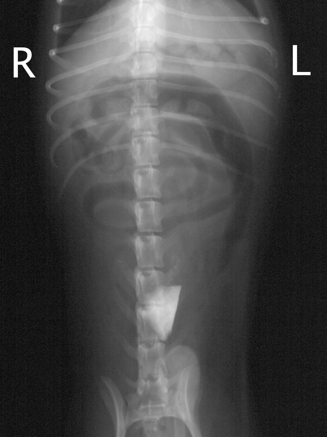
Figure 2 – Pneumocolon highlights the entire colon, demonstrating that the rock is in the small intestines.

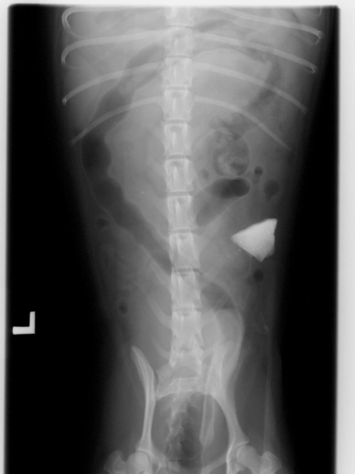
Figure 1 – Survey VD radiograph of a vomiting 2 yo Shih-tzu. A rock foreign body is visible in the mid-caudal abdomen.
Pneumocolon in the dog was described in 1978, and the clinical indications and technique remain essentially unchanged today.1 Most commonly, pneumocolon is indicated when a radiographically detectable foreign object is seen in the abdomen, but it is unclear whether the object is located in the small or large intestines. Pneumocolon can also be performed in cases where a single gas-dilated intestinal segment is seen, but it is unclear whether it represents a normal colon or abnormal small intestine. Repeat radiographs following insufflation of the colon with air will usually allow for definitive identification of the lesion in question.
The equipment required for a pneumocolon consists of a red rubber catheter, lubricant gel, and a large syringe. Sedation is usually unnecessary with reasonably cooperative patients. The red rubber catheter is introduced per rectum, and advanced as far as possible (ideally at least to the mid descending colon). Approximately 3-5 ml/kg of room air is then injected through the red rubber catheter, and abdominal radiographs are repeated. If an insufficient amount of air is seen within the colon, the procedure can be repeated. No complications from pneumocolon have been described.

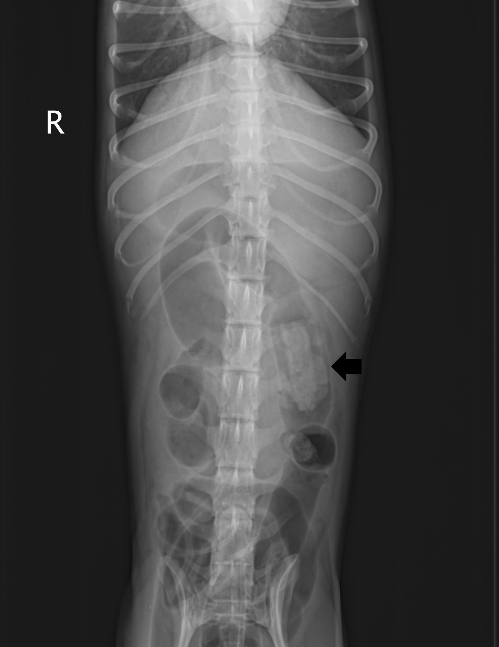
Figure 3 – Survey VD radiograph of a vomiting 10 yo poodle. A rectangular soft tissue foreign body is seen in the right mid-abdomen (arrow).
The colon is typically seen on a ventrodorsal abdominal radiograph with a “question mark” appearance. The cecum varies in size and shape from a short “C” to a spiral or corkscrew, typically is located in the mid to right side of the abdomen, and indicates the most proximal portion of the colon. The ascending colon extends cranial to the cecum, then turns to the left into the transverse colon. The transverse colon courses to the left just caudal to the stomach, then turns caudally into the descending colon. The descending colon extends caudally through the pelvic canal into the rectum. On a lateral view, the colonic segments are frequently superimposed with each other, which can make differentiation somewhat difficult. The descending colon is most readily identified on a lateral view, coursing just ventral to the kidneys and entering the pelvic canal dorsal to the urinary bladder.
Two case examples of pneumocolon are presented here. Orthogonal views both before and after pneumocolon were obtained, although only the most instructive views are shown. The first case is a 2 year old castrated male Shih-tzu with a history of vomiting. A large, angular mineral foreign object consistent with a rock is seen in the mid-caudal abdomen (Figure 1). Gas is seen in the transverse and proximal descending colon, although the colon is not identified at the level of the rock. Pneumocolon allows visualization of the entire colon and cecum, demonstrating that the rock is small intestinal in location (Figure 2). At surgery, the rock was located in the jejunum, with focal necrosis of the wall, necessitating a jejunal resection and anastomosis.
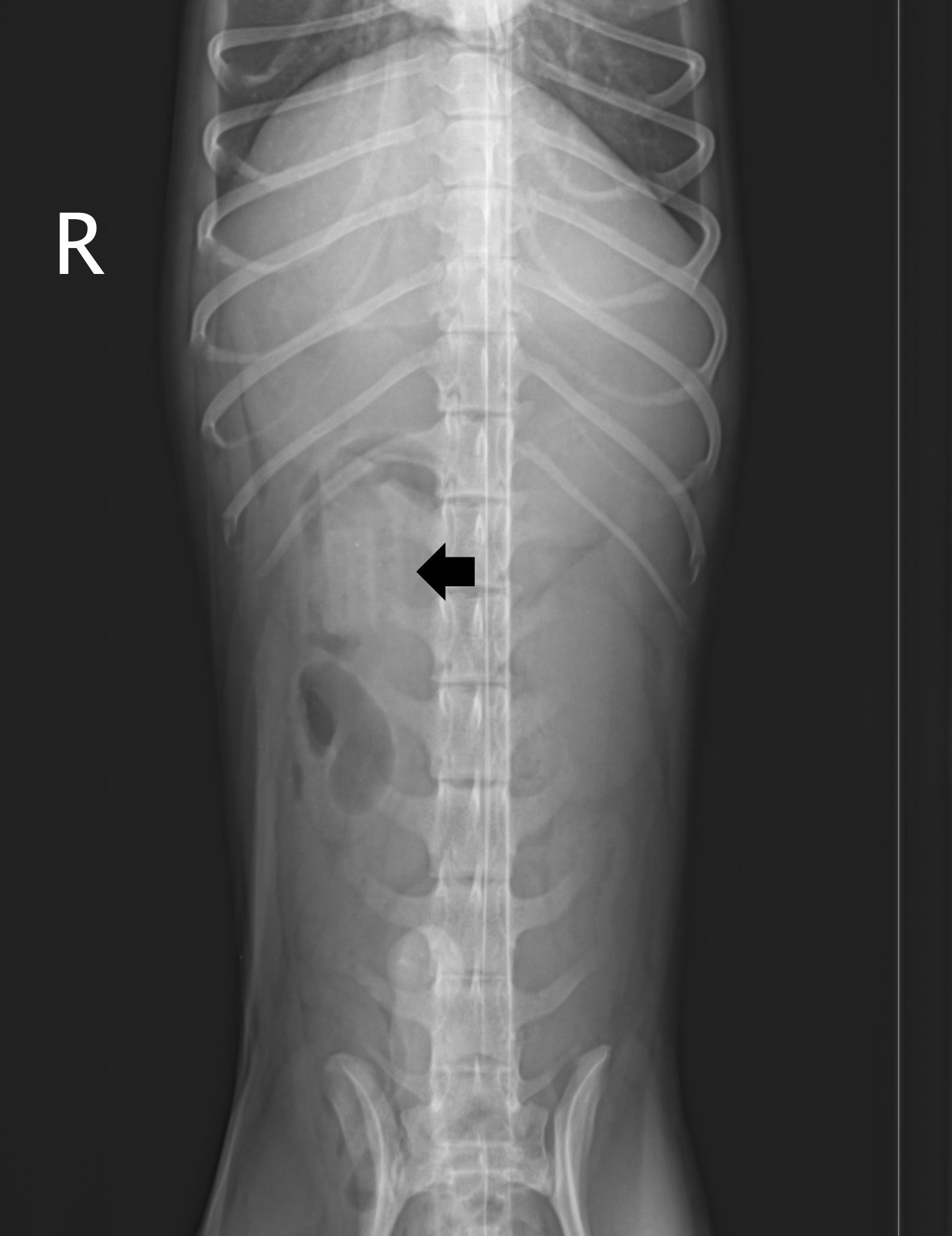
The second case is a 10-year-old spayed female miniature poodle with a history of vomiting. A rectangular soft tissue opacity is seen in the mid-abdomen on survey radiographs, and the colon is not readily identified (Figure 3). Pneumocolon helps to identify the foreign object within the lumen of the descending colon (Figure 4). Following subcutaneous fluid administration and observation, a rectangular plastic object was passed in the patient’s next bowel movement.
In summary, pneumocolon is a rapid, easy to perform, and clinically useful technique applicable in the ER or general practice setting to help differentiate surgical from non-surgical foreign bodies.
References
- Nyland TG, Ackerman N. Pneumocolon: a diagnostic aid in abdominal radiography. Vet Radiol 1978;19:203-7.
