-
Adopt
-
Veterinary Care
Services
Client Information
- What to Expect – Angell Boston
- Client Rights and Responsibilities
- Payments / Financial Assistance
- Pharmacy
- Client Policies
- Our Doctors
- Grief Support / Counseling
- Directions and Parking
- Helpful “How-to” Pet Care
Online Payments
Emergency: Boston
Emergency: Waltham
Poison Control Hotline
-
Programs & Resources
- Careers
-
Donate Now
 By Michele James, DVM, DACVIM(Neurology)
![]()
angell.org/neurology
neurology@angell.org
617-989-1666
The neurologic examination is the most important and arguably the most valuable diagnostic test in clinical neurology. It allows a clinician to not only determine if neurologic disease is present in a patient, but it also helps to localize where the lesion is anatomically and serves as a marker to assess improvement or progression of disease. With a thorough patient history, general physical examination, and neurologic examination, a clinician is equipped to form a list of differential diagnoses, form an appropriate diagnostic plan, and in some cases prioritize treatments. The neurologic examination requires very little instrumentation with only a light source, pleximeter, and hemostat needed.
The neurologic examination evaluates a patient’s mentation, posture and gait, cranial nerve function, conscious proprioception, spinal reflexes, pain, and nociception.
Mentation
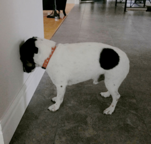
A patient’s mentation and level of consciousness should be observed before hands-on examination. This can be performed by allowing the patient to walk freely in an enclosed space or exam room. Mentation is generally described using the following qualifiers: Normal, Obtunded, Stupor, or Comatose. A patient with normal mentation is noted to be bright, alert, and responsive to their surround ings. An obtunded patient is noted to be dull and though conscious, may be less responsive to their surroundings. Stupor may be used to describe a somnolent patient whose level of consciousness is markedly decreased; however, the patient is responsive to noxious stimuli. Comatose is used to describe an unconscious patient that is not responsive to noxious stimuli. In addition to mentation, abnormal behaviors may also be observed, such as aggression, head pressing, and compulsive pacing or circling.
Posture and Gait
A patient’s posture and gait should also be observed before a hands-on examination, if possible. Evaluation of a pet’s posture at rest and gait changes can aid in neurolocalization. A pet exhibiting kyphosis of the thoracolumbar spine and low head carriage may have marked cervical pain. Gait changes may be secondary to paresis, ataxia, and pain. A clear description of the patient’s ambulatory status and degree of paresis, if present, should be noted. A patient is considered mobile if they can walk without assistance on all limbs. A patient is considered non-ambulatory if they cannot walk and support their weight without help. Paresis describes weakness and decreased voluntary motor function, whereas paralysis describes the absence of voluntary motor function.
Ataxia is primarily described in three forms: Proprioceptive, Cerebellar, or Vestibular. Proprioceptive ataxia is secondary to a lesion affecting the proprioceptive tracts extending from the brain down the length of the spinal cord. Proprioceptive ataxia may be characterized by scuffing or knuckling of the paws due to the patient not being fully aware of where their limbs are in space. Cerebellar ataxia is characterized by a hypermetric gait, truncal sway, and intention tremor. Cerebellar ataxia in a patient indicates a lesion affecting the spinocerebellar tracts. Vestibular ataxia is characterized by a patient veering to one side, falling to one side, and/or violently alligator rolling. Vestibular ataxia is secondary to a lesion affecting the vestibular system, and patients may also exhibit a head tilt, nystagmus, and a positional ventral strabismus ipsilateral to the head tilt.
A change in gait due to pain in the form of lameness may also be noted on initial observation. A thorough hands-on physical examination, including an orthopedic examination, can help determine if the lameness results from primary orthopedic disease versus root signature secondary to nerve impingement.
Cranial Nerve Function
Cranial nerve evaluation is accomplished by assessing a series of reflexes and responses and observation for facial symmetry. The presence of cranial nerve deficits can help with more specific neurolocalization of a lesion within the brain. The author typically begins her cranial nerve exam by assessing the eyes. The patient’s pupil size and symmetry and direct and consensual pupillary light reflexes are assessed in both eyes using a bright light source, such as a transilluminator (CN II, CN III). The menace response is then evaluated by covering one of the patient’s eyes and then making a menacing gesture with the clinician’s hand towards the uncovered eye and observing for a blink (CN II, CN VII). The menace response is learned and should be present in puppies and kittens by 12 weeks of age. The medial and lateral canthus of the eyes may then be gently palpated with the clinician’s finger or tip of the hemostat to assess for a blink (CN V, CN VII). The eyes should be observed for any strabismus or pathologic nystagmus (CN III, IV, VI, VIII). The facial sensation may be assessed by gently touching the patient along the muzzle and nasal mucosa within each nostril and observing the patient lift the lip and/or move their head away (CN V, CN VII). Facial symmetry should be noted by assessing for any atrophy of the muscles of mastication (CN V), lip droop or ear droop (CN VII), and head tilt (CN VIII). Lastly, a gag reflex may be assessed by opening the patient’s mouth and touching the tongue (CN V, CN IX, CN X, CN XII).
Conscious Proprioception
Conscious proprioception allows the patient to know where their limbs are in space and is controlled by the proprioceptive pathways that run from the brain down the length of the spinal cord. Delays in proprioception can be sensitive indicators of a neurologic lesion and, in conjunction with the remainder of the neurologic examination, can help determine neurolocaization and differential diagnoses. The author generally evaluates proprioception via paw placing. This assessment involves placing the pet in a standing position and, while supporting their weight, gently flipping over one paw so that it is knuckled on the ground and evaluating how quickly the pet corrects their paw. This test is repeated with each paw separately to evaluate all paws.
Spinal Reflexes
Spinal reflexes evaluate the sensory and motor components of the reflex arc. In conjunction with the remainder of the neurologic examination, spinal reflexes can help determine neurolocalization and differential diagnoses. The author finds that the most reliable and repeatable spinal reflexes to evaluate in the cat and dog are the patellar reflex, forelimb and hindlimb withdrawal reflex, and cutaneous trunci reflex. It should be noted that the patient should be relaxed to elicit a patellar reflex adequately.
Pain and Nociception
The last part of the neurologic examination is an assessment for spinal pain. The patient’s cervical and thoracolumbar spine should be palpated with increasing pressure to see if pain is elicited. The patient’s chest and abdomen should be supported during palpation. The range of motion of the neck should be evaluated. A treat may be used to encourage the pet to move their head side to side and up and down to assess lateral, dorsal, and ventral flexion of the neck.
Neurolocalization and Forming Differential Diagnoses
Using the information gathered during the neurologic examination, namely any neurologic deficits, the clinician can localize the patient’s lesion to the correct neuroanatomical location. These include Forebrain, Cerebellum, Brainstem, C1-5, C6-T2, T3-L3, L4-S, and Neuromuscular. Once the lesion is localized, the clinician can use this information with the pet’s clinical history to formulate a list of differential diagnoses and an appropriate diagnostic plan.
The chart below lists some of the more common differential diagnoses affecting the nervous system in dogs and cats:
| Cause | Example |
| Degenerative | • IVDD, Wobblers
• Degenerative Myelopathy, GOLPP • Storage Diseases |
| Anomalous (Congenital) | • Hydrocephalus
• Chiari malformation |
| Metabolic | • Hepatic/Renal Disease
• Electrolyte Disorders • Hypothyroidism |
| Neoplasia | • Meningioma, Glioma, PNST, etc. |
| Nutritional | • Thiamine Deficiency |
| Inflammatory | • MUE, SRMA |
| Infectious | • Discospondylitis, FIP, Distemper, Rabies |
| Idiopathic | • Idiopathic epilepsy
• Idiopathic head tremors |
| Trauma | • Head Trauma, Fracture |
| Toxin | • Lead, Bromethalin, Organophospahtes |
| Vascular | • Stroke, FCE |