-
Adopt
-
Veterinary Care
Services
Client Information
- What to Expect – Angell Boston
- Client Rights and Responsibilities
- Payments / Financial Assistance
- Pharmacy
- Client Policies
- Our Doctors
- Grief Support / Counseling
- Directions and Parking
- Helpful “How-to” Pet Care
Online Payments
Emergency: Boston
Emergency: Waltham
Poison Control Hotline
-
Programs & Resources
- Careers
-
Donate Now
 By Audrey Koid, DVM, DACVECC-SA
![]() angell.org/emergency
angell.org/emergency
emergency@angell.org
617-522-7282
March 2023
x
x
Respiratory distress is a common cause of an emergency visit, where prompt recognition and intervention could help save lives. Tools that can help with diagnosis include signalment, history, observation of breathing pattern, auscultation of the heart and lungs, and diagnostic imaging.
Respiratory Physiology
The respiratory system can be divided anatomically into the upper and lower airways, pulmonary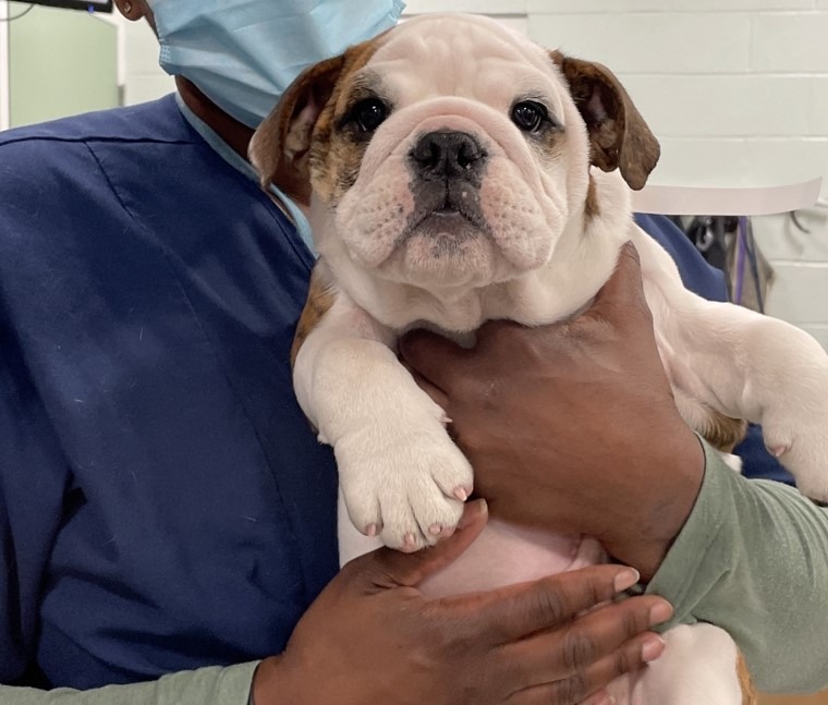 parenchyma, pleural space, and thoracic wall/diaphragm. The two main functions of the respiratory system are ventilation and oxygenation. Ventilation is the process of moving air in and out of the lungs. Once at the level of the alveoli, gas is exchanged between blood and air. Hypoxemia can occur due to problems with ventilation and/or gas exchange.
Normal breathing is carbon dioxide-driven. When PaCO2 rises, CO2 diffuses into the cerebrospinal fluid, liberating hydrogen ions that stimulate the central chemoreceptors to increase ventilation. This response is magnified if PaO2 is lowered. PaO2 becomes the primary driver of respiration when PaO2 drops below 50 mmHg at high altitudes and in some patients with lung disease (e.g., COPD).
Initial Assessment of a Patient in Respiratory Distress
 The age and breed of the patient are great places to start to narrow down the possible causes of respiratory distress. Younger patients are more likely to have something infectious, trauma, or congenital-related, while neoplasia and degenerative conditions are much more likely in older animals. Most, if not all of us, automatically think that a loudly breathing brachycephalic breed has an upper airway obstruction, predisposing them to heat stress/stroke. This “pattern recognition” is helpful to allow for quick intervention. Returning to the example of the brachycephalic dog with likely upper airway obstruction, sedation, and potential intubation will make more of a difference than oxygen therapy alone.
Next, observe the patient. Is there loud or noisy breathing usually indicating an upper airway issue? Are there any signs of trauma or abdominal distension? What type of breathing pattern is the patient showing? A restrictive breathing pattern characterized by rapid and shallow breaths in a cat is likely to have pleural effusion. In contrast, a cat with an expiratory effort is likelier to have asthma.
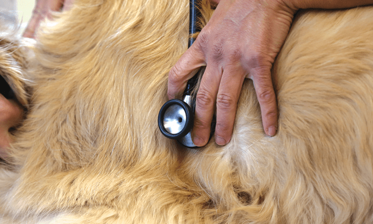
A heart murmur, gallop sounds, pulmonary crackles, or wheezes could narrow your differential further when auscultating patients. If the lung or heart sounds are muffled, there is a good chance of pleural space disease.
Temperature is sometimes overlooked as a tool to narrow down the differential list. A hyperthermic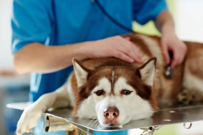 animal is more likely to have an infection (pneumonia or pyothorax) or an upper airway obstruction. In contrast, a hypothermic animal is usually in shock and is commonly seen in cats with congestive heart failure.
Initial Stabilization of a Patient in Respiratory Distress
Oxygen therapy is helpful in all cases but more helpful in some than others, as mentioned with the brachycephalic dog with upper airway obstruction. Another situation where oxygen support provides less benefit is in patients with pleural space disease, where a therapeutic thoracocentesis to remove fluid or air allows for better expansion of the lungs with very noticeable improvement in the patient’s breathing.
Respiratory distress is reportedly panic-inducing in people, so sedation could help the patient calm down and decrease the work of breathing. On the flip side, the sedation may depress the patient’s respiratory drive, which could lead to worse hypoxia. Two general rules of thumb are to avoid sedating animals that appear very tired but catch themselves right before they fall asleep and be prepared to intubate any animal that has been given a sedative. It would also be ideal to have intravenous access for quicker administration of medications, especially if the patient requires intubation. Still, the placement of the IV catheter should be weighed against the stress of restraint.
More definitive treatment would depend on the cause of respiratory distress, but the following drugs are useful to have on hand and can be administered on triage:
- Sedatives:
a. Butorphanol 0.1–0.4 mg/kg IV or IM
b. Acepromazine 0.005 – 0.02 mg/kg IV or IM - Furosemide 1–2 mg/kg IV or IM for patients with suspected congestive heart failure
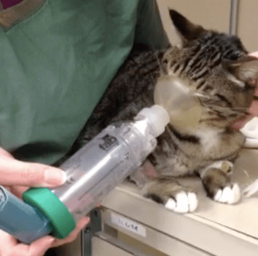
- Albuterol 1–2 puffs/cat if asthma is suspected
- Terbutaline 0.01 mg/kg IM or SQ for asthma or chronic bronchitis
Next Steps
Once the patient is more stable, additional diagnostics such as radiographs, point-of-care ultrasound, lab work, and sedated oral exams can be performed to confirm the cause of respiratory distress. If the cause is unclear in a markedly dyspneic animal based on initial assessment, point-of-care ultrasound may help narrow down the options as it can be done quickly either through the window of the oxygen cage or with a helper administering flow-by oxygen to the patient.
 The response to your initial stabilization, if your educated guess as to the cause of respiratory distress is correct, will generally result in a noticeable improvement in the patient fairly quickly. If you do not see this improvement, you will need to reassess your patient to determine whether they require an escalation of interventions for that particular disease process or whether a different or concurrent disease process is going on. For example, a brachycephalic animal with upper airway obstruction may require more sedatives than what was initially given on triage to calm them down and may also have concurrent aspiration pneumonia. Some brachycephalics may require heavy sedation and intubation for several hours (or longer) to decrease the inflammation in their upper airway enough to be safely extubated.
If any animal appears to be in imminent danger of respiratory fatigue, they should be intubated (and ideally mechanically ventilated) while a more definitive plan is made, even if the ultimate decision is euthanasia. Our goal is to always prevent suffering in the patients we treat.
Conclusions
As the name indicates, respiratory distress can be stressful for all parties involved, especially given its dynamic nature. However, using a systematic approach and the tools that are available at most, if not all, veterinary hospitals, from pattern recognition and situational awareness to good history taking and physical examination to more specific diagnostics such as point-of-care ultrasound and thoracic radiographs, acting quickly to stabilize these patients can make a big difference, and once definitive treatment is implemented, most animals do recover from their respiratory crisis.
x
References
- West J. B. & Luks A. (2016). West’s respiratory physiology: the essentials (Tenth). Wolters Kluwer.
- Drobatz, K. J., Hopper, K., Rozanski, E. A., & Silverstein, D. C. (Eds.). (2018). Textbook of small animal emergency medicine. John Wiley & Sons.
- Silverstein D. C. & Hopper K. (2015). Small animal critical care medicine (Second). Elsevier/Saunders.
- Tong CW, Gonzalez AL. Respiratory Emergencies. Vet Clin North Am Small Anim Pract. 2020 Nov;50(6):1237-1259. doi: 10.1016/j.cvsm.2020.07.002. Epub 2020 Sep 2. PMID: 32891440.
- Sumner C, Rozanski E. Management of respiratory emergencies in small animals. Vet Clin North Am Small Anim Pract. 2013 Jul;43(4):799-815. doi: 10.1016/j.cvsm.2013.03.005. Epub 2013 Apr 20. PMID: 23747261.
- Lisciandro GR, Lisciandro SC. Lung Ultrasound Fundamentals, “Wet Versus Dry” Lung, Signs of Consolidation in Dogs and Cats. Vet Clin North Am Small Anim Pract. 2021 Nov;51(6):1125-1140. doi: 10.1016/j.cvsm.2021.07.012. Epub 2021 Sep 14. PMID: 34535335.
- Lemieux E, Rozanski E, Buckley G, Chalifoux N, Kennedy C, Lynch A, Rutter C, Tracy A, Silverstein DC. Indications and outcomes for puppies undergoing mechanical ventilation: 59 cases (2006 to 2020). Can Vet J. 2021 Aug;62(8):839-842. PMID: 34341595; PMCID: PMC8281947.