-
Adopt
-
Veterinary Care
Services
Client Information
- What to Expect – Angell Boston
- Client Rights and Responsibilities
- Payments / Financial Assistance
- Pharmacy
- Client Policies
- Our Doctors
- Grief Support / Counseling
- Directions and Parking
- Helpful “How-to” Pet Care
Online Payments
Referrals
- Referral Forms/Contact
- Direct Connect
- Referring Veterinarian Portal
- Clinical Articles
- Partners in Care Newsletter
CE, Internships & Alumni Info
CE Seminar Schedule
Emergency: Boston
Emergency: Waltham
Poison Control Hotline
-
Programs & Resources
- Careers
-
Donate Now
 By Michael M. Pavletic, DVM, DACVS
![]()
angell.org/surgery
surgery@angell.org
617-541-5048
January 2022
Introduction
Tail fold pyoderma is frequently associated with the deviated tails most commonly found in brachycephalic breeds. Most cases are in English bulldogs. This condition is less commonly seen in French bulldogs, Boston terriers, and pugs. The colloquial expression for this condition is “screw tail,” since the spiral of the tail resembles a corkscrew. It is less commonly called “ingrown tail.”
The abnormal shape of the terminal coccygeal vertebrae can result in the inability to lift the tail manually. In many bulldogs, this tail deviation may have a limited effect on the dog, with little or no dermatitis associated with the tail folds. In others, the underlying base of the tail may form a deep crease or depression that extends 3 to 5 centimeters cranially. A cotton-tipped applicator can be used to probe the depth of this skin pocket. The abnormal tail can also take a ventral twist or bend in some bulldogs, covering the anus. In more severe cases, the dog is forced to defecate with the stool being extruded ventral and lateral to this tail obstruction.
Although deeply creased dorsal skin folds may develop dermatitis (intertrigo), the primary problem area is the ventral crease beneath the tail base. This area accumulates moisture, and the dense hair growth in this cleft creates the ideal condition for chronic dermatitis. It is common for a foul odor to form due to this condition. Fecal contamination also can precipitate infection.
Owners may clean this tail base area with medicinal wipes, such as Duoxo 3% Chlorhexidine Pads. Cleaning with wipes may help the patient in mild cases, but the condition often progresses. The patient will scoot or rub his rear area on the carpeting in the house to relieve the itch and discomfort. Some bulldogs become resistant to anyone approaching the rear area and will attempt to bite. In these problematic cases, amputation of the tail and the ventral skin fold excision is required. Successful surgical resolution of screw tail will eliminate the itch and pain: in many cases, the dog is calmer, and the canine aggression to “protect” his rear end is eliminated.
Surgical Technique
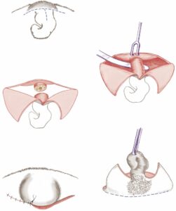
Surgery is primarily directed at (1) resecting the abnormal terminal coccygeal vertebrae, cranial to the major kind or bent of the tail, and (2) removing the ventral crease or fold. The earliest technique reported the veterinary advocated the removal of the entire tail skin and underlying deviated tail using a broad elliptical incision. Resection and closure result in a dog with a comparatively flat surface area. The author’s technique is a more cosmetic option by preserving a major dorsal skin fold to form the semblance of a short tail upon closure (Figure 1 & 2).
Comments and Caveats
During amputation, care is taken to avoid deeper dissection that may perforate the dorsal rectal wall. The dorsal portion of the external anal sphincter is safely distal to the surgical incisions. The most challenging part of the surgery is in those cases where the dorsal dermal surface of the recessed skin pocket is fibrosed to the undersurface of the coccygeal vertebrae. Dissection of the skin from the ventral vertebral surface requires patience to ensure it is removed in its entirety. Leaving a segment of skin would eventually create a foreign body reaction in response to the growth of skin epithelium and the associated hair follicles. I use absorbable Monocryl (Ethicon, Polyglicaprone) for external sutures: Monocryl sutures typically degrade and fall out of the skin around four weeks after surgery.
Pain Management and Postoperative Care
An epidural is customarily performed before surgery. Nocita (Elanco, bupivacaine liposome injectable suspension) is infused subcutaneously at the surgical site at the time of surgical closure. The patient is typically sent home on a broad-spectrum antibiotic (normally Clavamox [Zoetis, amoxicillin, and clavulanate potassium tablets]) for approximately five days and Gabapentin (Neurontin). No stool softeners are needed. Patient exercise is restricted for a minimum of one week before reintroducing the dog to normal activities.
Legend
Figure 1: Surgical Technique
(A) A “T” incision is created caudal to the cranial skin fold at the base of the tail (dashed lines).
(B) The triangular skin flaps created by the T incision are retracted, and dissection is directed to exposure of the coccygeal vertebrae anterior to the point of tail deviation. A Carmalt clamp can be directed beneath the tail to protect the underlying dorsal rectal wall.

Figure 1
(C) Electrocautery is typically used to control bleeding vessels: vascular clips or ligatures may be used for larger vessels. Bone cutters are then cut directly through or between the exposed vertebra. [Alternatively, some surgeons will use a scalpel to divide between individual coccygeal vertebrae. Other surgeons will pass a Gigli wire to severe the tail.]
(D) With the successful division of the vertebra, the loose terminal tail segment is grasped with forceps. Firm traction is applied in a dorsal direction; this facilitates eversion of the invaginated or recessed ventral cutaneous fold. A curved incision is made at the ventral base of the tail fold (dashed line), completing the resection of the tail and ventral skin crease. Metzenbaum scissors can be used to undermine and mobilize the ventral skin fold.
(E) After resectioning the tail and associated ventral skin folds, the area is lavaged and examined for any remaining bleeding vessels before closure. Absorbable sutures may be used to close dead space before approximating the skin margins with absorbable interrupted intradermal sutures. Simple interrupted skin sutures are used to complete the skin closure. The large preserved cranial skin fold forms a prominent peak, giving the semblance of a short, symmetrical tail.
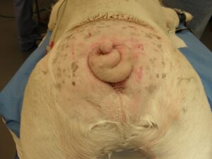
Figure 2A

Unusual tail deviation in the bulldog leaves a prominent dorsal skin ridge on the right side (Figure 2A, above).
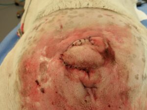
Figure 2B

Figure 2B
The deviated coccygeal vertebrae were severed with bone cutters. A modified curved skin incision was created to preserve the large dorsal skin fold. The salvaged skin fold created the impression of a small tail.
References
- Pavletic MM. Miscellaneous Reconstructive Surgical Techniques. In: Atlas of Small Animal Wound Management and Reconstructive Surgery, 4th ed. Hoboken: Wiley Blackwell, 2018; 823-826, 842-3.
- (Pavletic MM. Miscellaneous Reconstructive Surgical Techniques. In: Atlas of Small Animal Wound Management and Reconstructive Surgery, 4th ed. Hoboken: Wiley Blackwell, 2018; 826.)