-
Adopt
-
Veterinary Care
Services
Client Information
- What to Expect – Angell Boston
- Client Rights and Responsibilities
- Payments / Financial Assistance
- Pharmacy
- Client Policies
- Our Doctors
- Grief Support / Counseling
- Directions and Parking
- Helpful “How-to” Pet Care
Online Payments
Referrals
- Referral Forms/Contact
- Direct Connect
- Referring Veterinarian Portal
- Clinical Articles
- Partners in Care Newsletter
CE, Internships & Alumni Info
CE Seminar Schedule
Emergency: Boston
Emergency: Waltham
Poison Control Hotline
-
Programs & Resources
- Careers
-
Donate Now
By Zachary Crouse, DVM, DACVIM (SAIM)![]()
angell.org/internalmedicine
internalmedicine@angell.org
617-522-7282
May 2023
XX
XX
Introduction
Chronic cough in dogs is defined as coughing on most days for a period of greater than eight weeks. Common differential diagnoses for a chronic cough include airway collapse, infectious disease (bacterial, fungal, and parasitic), neoplasia, interstitial lung disease, eosinophilic bronchopneumopathy, and chronic bronchitis. Bronchoscopy is an underutilized but beneficial tool in diagnosing and developing a treatment plan for dogs with a chronic cough.
When should bronchoscopy be considered?
The typical workup for chronic cough begins with a minimum database and thoracic radiographs. CBC and chemistry values are often not specific to a particular diagnosis but may show signs of inflammation or elucidate other comorbidities that could be important for the treatment plan. Thoracic radiographs aid in the exclusion of neoplasia, pleural effusion, and cardiac disease. For dogs that do not have a clear radiographic diagnosis, bronchoscopy is often the next step. Almost all dogs with more than two months of cough will have abnormalities on bronchoscopic examination. Bronchoalveolar lavage, performed as part of a standard bronchoscopy, also allows for cytology and culture of the lower airways.
Bronchoscopy
Bronchoscopy includes evaluating the lower airways for collapse, hyperemia, thickened or irregular mucosa, and mucus. Below are examples of bronchoscopic images obtained in common lower airway diseases.
X
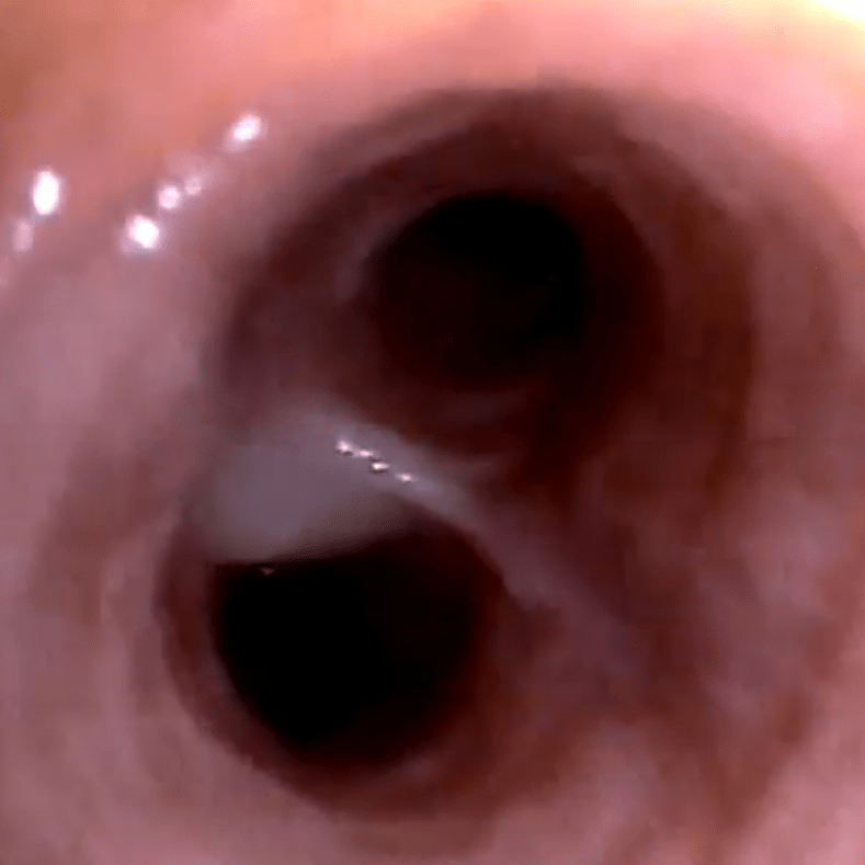
Figures 1 and 2 (top and bottom): Images from a 10-year-old Labrador retriever with chronic bronchitis. Note the increased mucus, hyperemic and thickened bronchial mucosa, and slightly nodular appearance. Chronic bronchitis is diagnosed by exclusion of other chronic lower airway disease. Almost all dogs with chronic bronchitis will have abnormalities on bronchoscopy.
X
XXX
XXX
XX
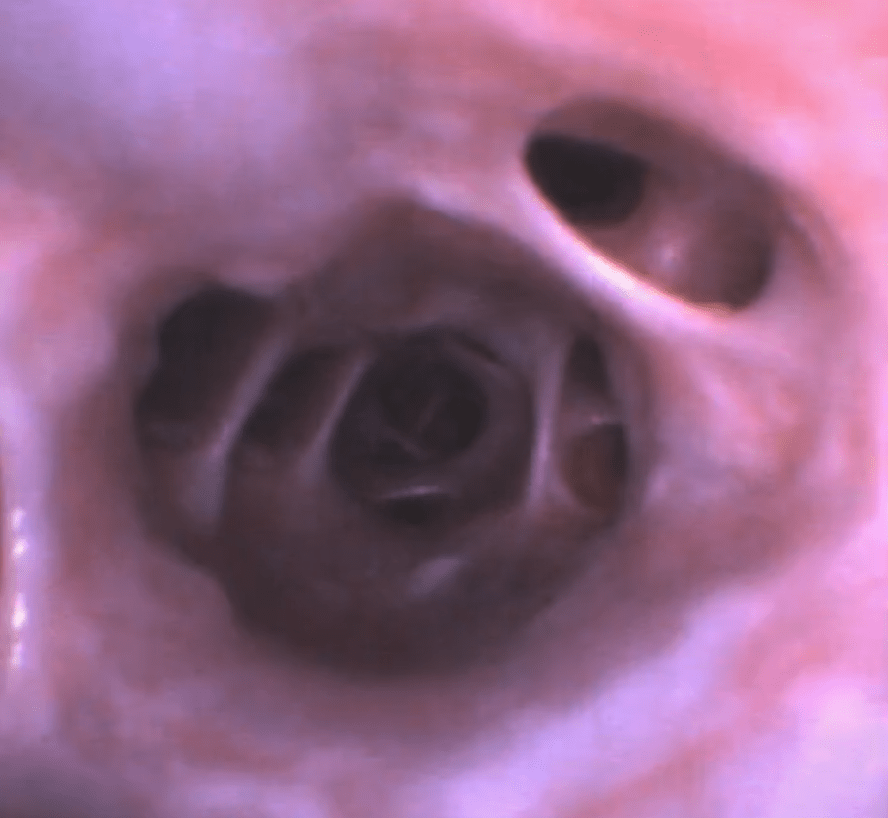
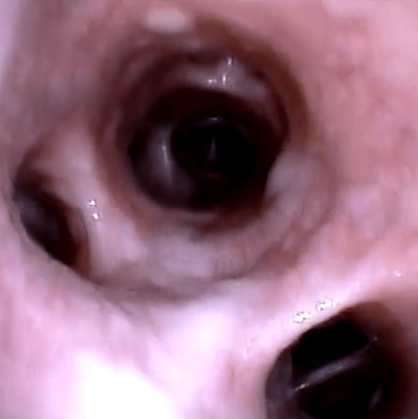
Figure 3: Image from an 8-year-old mixed breed dog demonstrating bronchiectasis. Bronchiectasis occurs secondary to an inflammatory or infectious insult. Studies have shown bronchoscopy to be a highly effective means of identifying cases of bronchiectasis and will often also diagnose the primary underlying pathology leading to bronchiectasis.
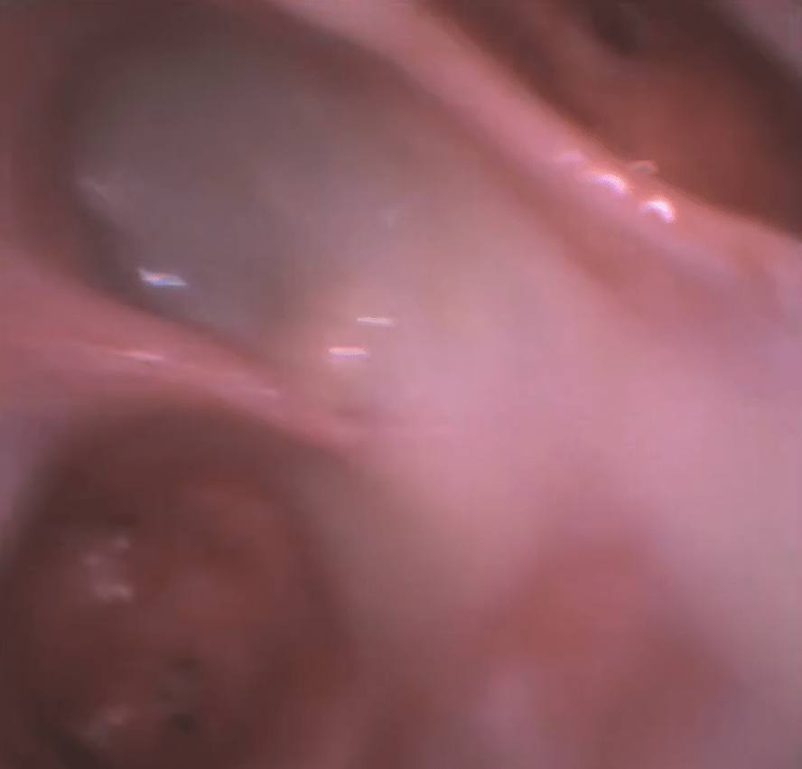
Figure 4: Image from a 6-year-old mixed breed dog with chronic bronchitis that has progressed to saccular bronchiectasis and secondary bacterial pneumonia. Note the purulent material pooled in the bronchi.
XXX
XXX
XX
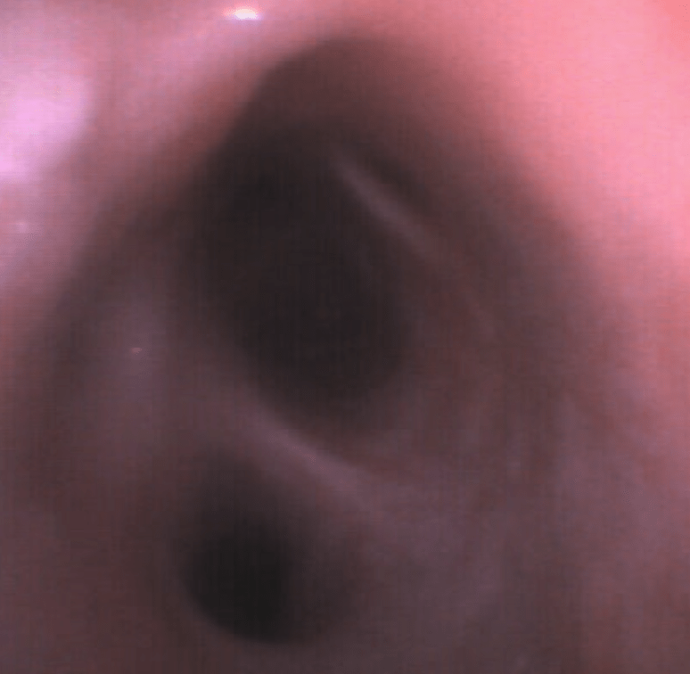
Figures 5 and 6 (top and bottom): Image from a 13-year-old Miniature Poodle demonstrating lower airway collapse. The image on the left is the airway during inspiration, and the image on the right demonstrates dynamic collapse during expiration. Approximately half of all dogs presenting with a chronic cough will have tracheobronchomalacia and secondary airway collapse, which may occur with other concurrent bronchial disease.

X
Bronchoalveolar lavage
Bronchoalveolar lavage (BAL) involves instilling small aliquots (typically 3 to 10 ml, depending on the size of the patient) of sterile saline through the bronchoscope and retrieving the majority of the sample into a sterile container using suction. Bronchoscopy-guided BAL allows a distinct advantage over a blind technique in that the saline can be directed to affected areas, and retrieval of the saline can be observed. BAL fluid is immediately submitted for cytology, culture, and infectious disease testing as appropriate to the individual case.
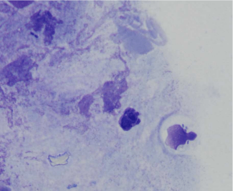
Figures 7 and 8 (top and bottom): Cytology images from a 13-year-old Pug with chronic bronchitis and bacterial bronchopneumonia. Note the phagocytized bacteria within the neutrophils. Bacterial infection is a common cause of acute worsening of chronic cough in dogs with chronic bronchitis. The chronic inflammation and secondary mucosal changes associated with chronic bronchitis may predispose to bacterial infection. Photo credit – Patty Ewing.
XXX
XXX
XX

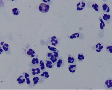
Figure 9: Cytology from a 10-year old-mixed breed dog with chronic cough. Note the lungworm larvae present in this sample. Eosinophilic inflammation may be seen with parasitic infection, allergic disease, or eosinophilic bronchopneumopathy, among others.
X
Conclusion
Many dogs with chronic cough have more than one underlying cause (i.e., airway collapse and chronic bronchitis or chronic bronchitis and bacterial bronchopneumonia), and it can be difficult to treat these patients with only a presumptive diagnosis appropriately. Bronchoscopy and bronchoalveolar lavage are useful tools that can often provide a definitive diagnosis. After the diagnosis has been made, therapy can be tailored appropriately. This often means treating an acute and chronic component of the patient’s disease.
x
x
References
Hawkins EC, Clay LD, Bradley JM, Davidian M. Demographic and historical findings, including exposure to environmental tobacco smoke, in dogs with chronic cough. J Vet Intern Med. 2010;24:825–831.
Johnson LR, Johnson EG, Vernau W, Kass PH, Byrne BA. Bronchoscopy, imaging, and concurrent diseases in dogs with bronchiectasis: (2003–2014). J Vet Intern Med. 2016;30(1):247–254
Johnson LR, Queen EV, Vernau W, et al. Microbiologic and cytologic assessment of bronchoalveolar lavage fluid from dogs with lower respiratory tract nfection:105 cases (2001-2011). J Vet Intern Med 2013;27:259–67.
Padrid PA, Hornof WJ, Kurpershoek CJ, et al. Canine chronic bronchitis: a pathophysiologic evaluation of 18 cases. J Vet Intern Med 1990;4:172–80
Rozanski E. Canine chronic bronchitis. Vet Clin North Am Small Anim Pract. 2014;44:107–116