-
Adopt
-
Veterinary Care
Services
Client Information
- What to Expect – Angell Boston
- Client Rights and Responsibilities
- Payments / Financial Assistance
- Pharmacy
- Client Policies
- Our Doctors
- Grief Support / Counseling
- Directions and Parking
- Helpful “How-to” Pet Care
Online Payments
Emergency: Boston
Emergency: Waltham
Poison Control Hotline
-
Programs & Resources
- Careers
-
Donate Now
 Michael M. Pavletic, DVM, DACVS
Director of Surgery
angell.org/surgery
surgery@angell.org
617-541-5048
Although relatively uncommon, the urine stream in male dogs can strike a portion of their body: this usually involves the forelimb or the anterior abdomen/caudal thoracic area. The undesirable consequence is the need for the pet owner to wash the area off after every urination. A urine odor normally persists with the dog. This condition, as well as its correction, was first reported by the author in 2009.
Since this article, the author has operated on a number of these patients. There are three general causes that I identified which contribute to a “misguided” urine stream listed below. How the patient stands during urination (four-legged vs. lifting a rear leg) also will influence the direction of the urine stream to a variable degree in association with the following causes.
(1) The body conformation of the dog may predispose the pet to this condition. Patients with a tucked abdomen and prominent “dome shaped’ chest can result in the urine stream striking the xyphoid area or lower forelimb. The angle of urination also is influenced by the natural length of the dorsal preputial skin fold reflecting off the caudal abdomen. This will influence the downward angle of the prepuce and the urine stream exiting the preputial ostium.
(2) A second cause of this condition is the result of surgery involving the preputial area. Typically, this is associated with tumor resection with subsequent closure of the cutaneous wound under tension. Postoperative incisional tension, in turn, can alter the angle of the prepuce as well as the position of the ostium.
(3) The “Preputial Advancement Technique,” to correct paraphimosis, can alter the direction of the urine stream postoperatively. In these patients, the prepuce is stretched during its forward advancement to cover the exposed penis. The preputial ostium is now in a more rostral position and the angle of urination can be altered to a variable degree. In affected dogs, the urine stream exits in a straight line towards the anterior abdomen and caudal thoracic area.
The preputial ostium has a major influence on the urine stream during micturition. Similarly the surgical modification of this orifice can be used to redirect the urine stream. The author’s technique involves closure of the dorsal half of the preputial ostium to create a deflective barrier which diverts the urine stream in a ventral or downwards direction (Figure 1).
Absorbable 3-0 Monocryl sutures (polyglecaprone) are typically used on the skin. Sutures normally degrade and fall out of the skin around four weeks after surgery.
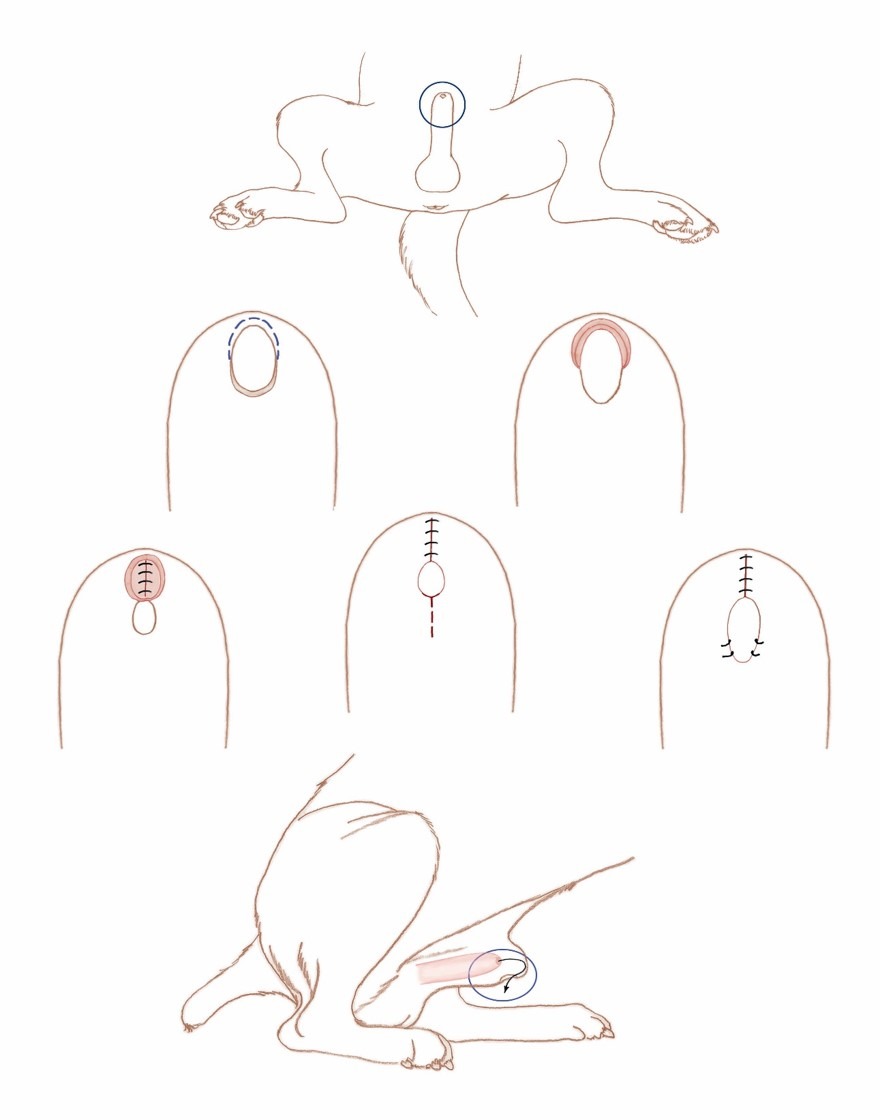 Figure 1. URINE DIVERSION TECHNIQUE
(A.) Focus on the preputial ostium.
(B.) The mucocutaneous junction of the dorsal half of the preputial ostium is trimmed or incised (dashed line) thereby separating the mucosal and cutaneous margins.
(C.) Complete separation of the mucosal and cutaneous surfaces.
(D.) 3-0 or 4-0 absorbable sutures are used to appose the mucosal margins.
(E.) The incised skin margins are apposed, completing the dorsal closure. The dashed line denotes the location to incise the ventral margin of the ostium.
(F.) The ventral incision is extended to create the original diameter of the ostium. The cutaneous margins are sutured to the opposing mucosal margins. The penis is extruded to assure it is capable or spontaneously sliding back into the preputial cavity prior to anesthetic recovery.
(G.) Transparent view of the penis within the prepuce. Urine exiting the penis strikes the preputial closure, deviating the urine stream ventrally (arrow).
(From Pavletic MM. Preputial Reconstructive Surgery. In: Atlas of Small Animal Wound Management and Reconstructive Surgery, 4th ed. Hoboken: Wiley Blackwell, 2018; 750.)
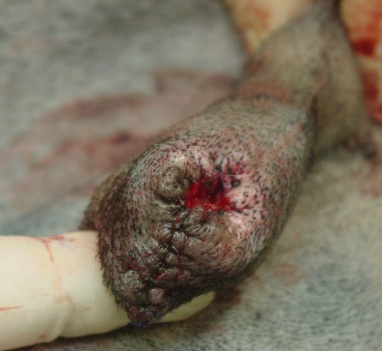 Figure 2. Postoperative view of closure of the dorsal preputial ostium followed by the reciprocal enlargement of the ventral preputial margin. The penis could be easily extruded through the modified ostium with its spontaneous retraction back into the preputial cavity. The urine stream, exiting the penis, strikes the dorsal ostium barrier created, diverting the stream ventrally between the patient’s limbs. (From Pavletic MM, Brum DE. (2009). Diversion of the urine stream by surgical modification of the preputial ostium in a dog. J AM Vet Med Assoc 235: 1067-1068.)
References
- Michael M. Pavletic, DVM, DACVS; Douglas E. Brum, DVM. Diversion of urine stream by surgical modification of the preputial ostium in a dog. J Am Vet Med Assoc 235:1067-1068, 2009.
- Pavletic MM. Preputial Reconstructive Surgery. In: Atlas of Small Animal Wound Management and Reconstructive Surgery, 4th ed. Hoboken: Wiley Blackwell, 2018; 731-771.