-
Adopt
-
Veterinary Care
Services
Client Information
- What to Expect – Angell Boston
- Client Rights and Responsibilities
- Payments / Financial Assistance
- Pharmacy
- Client Policies
- Our Doctors
- Grief Support / Counseling
- Directions and Parking
- Helpful “How-to” Pet Care
Online Payments
Emergency: Boston
Emergency: Waltham
Poison Control Hotline
-
Programs & Resources
- Careers
-
Donate Now
By Kristine Burgess, DVM, DACVIM (Medical Oncology)
angell.org/oncology
oncology@angell.org
617-541-5136
June 2024
x
Introduction

In human medicine, early cancer detection is well-established with multiple screening methods (e.g., mammography, colonoscopy) and newer approaches like blood-based multicancer early detection (MCED) testing. Conversely, in veterinary medicine, a cancer diagnosis is often limited to an annual or semiannual wellness visit with routine diagnostics tests, including a history, physical examination, and possibly basic tests (CBC, biochemistry, urinalysis). This can result in a late-stage diagnosis, leading to worse outcomes for our patients.
Early Detection of Canine Cancer
Several studies in the veterinary literature have identified specific mutations in a patient’s cancer, which can enhance early cancer detection and improve outcomes. They could also be integrated into routine veterinary care, potentially leading to earlier and more accurate cancer diagnoses and personalized care.
BRAF for Urogenital Tumors
A well-established example of cancer diagnosis/screening includes the molecular detection of a genetic alteration of the BRAF gene in voided urine. This is a non-invasive screening test for canine urogenital cancer. Cadet BRAFTM is a PCR-based genetic test that allows the detection of a mutation in the urothelial cells shed in the urine. The BRAF test allows preclinical detection in ~80% of dogs with bladder or prostate cancer before the development of overt cancer symptoms. This commercially available test has revolutionized the ability to diagnose a historically challenging cancer. The test is highly sensitive and beneficial for early diagnosis and may be useful in monitoring for cancer progression or regression. It is commercially available for both veterinarians and pet owners and is particularly useful as a screening test for certain breeds at risk.
Liquid Biopsy
Several non-standardized forms of non-invasive blood-based cancer screening tools have been developed and applied to a few different canine cancers, referred to as a “liquid biopsy.” These tests detect cancer-associated biomarkers or genomic mutations in a dog’s blood. This enables genomic profiling of cancer cases that are difficult to biopsy or diagnose in an early cancer stage. Most liquid biopsy tests available in veterinary medicine are promoted as multi-cancer early detection tests that can be used at regular intervals (e.g., annually) or cancer screening in high-risk populations.
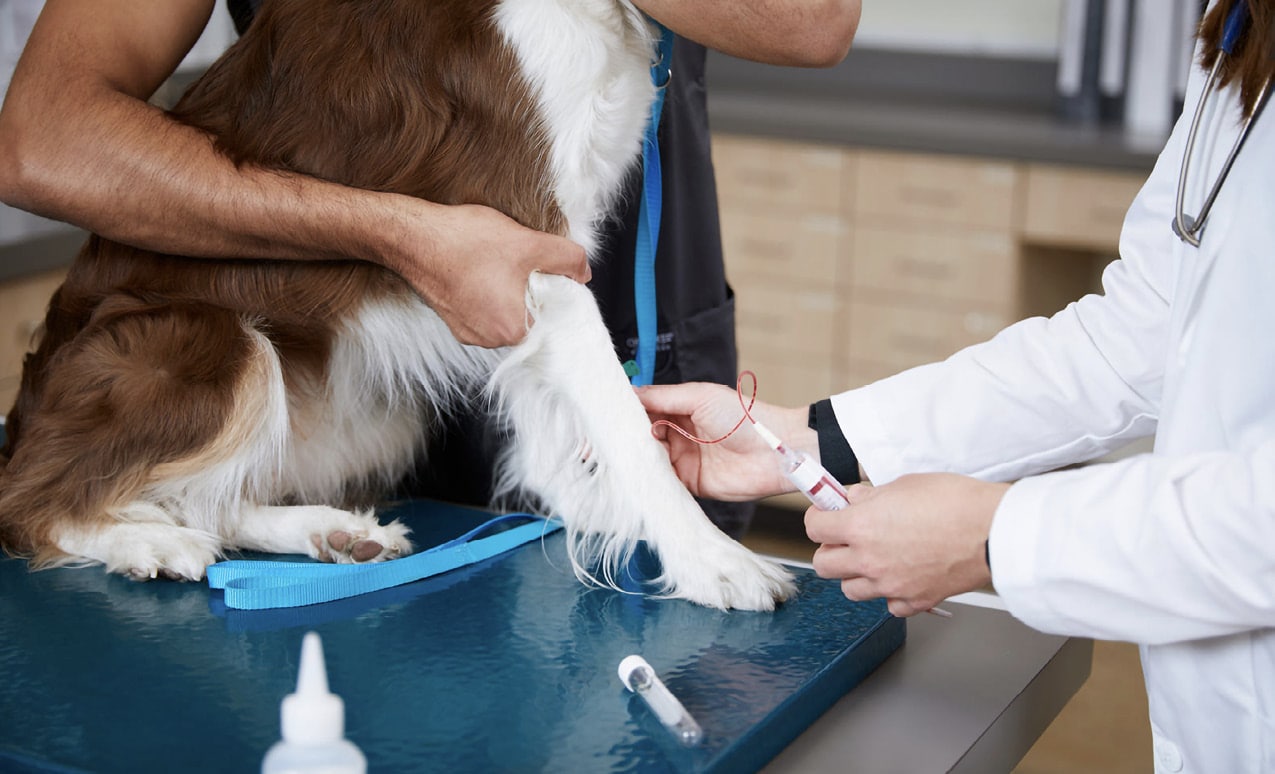 Currently, a few screening tests are available in the veterinary market, but not all liquid biopsy tests report the same information. Clinicians must know that commercially available veterinary liquid biopsy tests do not replace tissue-based diagnoses. Instead, they provide a non-invasive alternate method for cancer screening and aid in diagnosis and monitoring. These tests complement information gathered from the physical examination, clinical history, and other diagnostic tests.
In conclusion, liquid or blood-based biopsy tests represent a significant advancement in human and veterinary oncology by providing a non-invasive and comprehensive cancer diagnosis and management method.
Takeaways
- Liquid biopsy tests can detect specific cancer biomarkers in routine blood or urine samples.
- Although false-positive and false-negative results are possible, these tests can aid in diagnosing and monitoring cancer in dogs, and sensitivity/ specificity can vary. Therefore, results need to be interpreted with caution.
- A positive test should always be confirmed via tissue biopsy/cytology or other proven methods.
- Further research and clinical experience are warranted.
Advanced Imaging Techniques and Application in Practice
PET/CT
The integration of Positron Emission Tomography (PET) with Computed Tomography (CT) combines the strengths of both modalities to provide a comprehensive view of the cancer patient.
PET Imaging
- Biochemical and Physiological Insights: PET scans utilize radiotracers, such as Fluorodeoxyglucose (FDG), which mimics glucose in the body. This exploits the high glucose metabolism rates displayed by most cancer cells and allows for the visualization of metabolic activity, highlighting areas of increased uptake indicative of malignancy.
- Early Detection and Characterization: PET can detect biochemical changes before structural changes become apparent, enabling earlier diagnosis than CT or MRI.
CT Imaging
- Anatomic Detail: CT scans provide detailed images of the body’s internal structure. They excel in visualizing the size, shape, and precise location of lesions or abnormalities.
- Guidance for Biopsy and Surgery: The anatomic detail from CT scans assists in planning surgical interventions and guiding biopsies, ensuring precise targeting of areas of concern.
Combined PET/CT
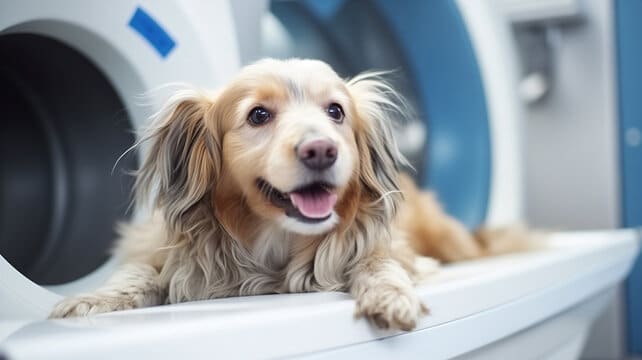
- Enhanced Diagnostic Accuracy: By combining metabolic information from PET with the anatomic detail from CT, PET/CT provides a more accurate diagnosis and staging of cancer. This hybrid approach reduces false positives and negatives, offering a clearer distinction between benign and malignant lesions.
- Improved Tumor Localization and Assessment: PET/CT allows for the precise localization of tumors and assessment of their metabolic activity. This is crucial for accurate staging, treatment planning, and monitoring of therapeutic response.
- Comprehensive Evaluation: The combined modality provides a one-stop imaging solution, reducing the need for multiple separate imaging tests. This leads to more efficient patient management and streamlines workflows in clinical practice.
Oncologic Applications of PET/CT
- Diagnosis and Initial Staging: PET/CT is invaluable in diagnosing and staging various cancers in humans. It helps determine the extent of disease spread, guiding treatment decisions.
- Therapeutic Monitoring: During and after treatment, PET/CT assesses the effectiveness of therapy by evaluating changes in metabolic activity and tumor size. This helps in adapting treatment plans based on the response.
- Detection of Recurrence: PET/CT effectively detects cancer recurrence, often identifying recurrent disease earlier than conventional imaging methods.
PET/CT represents a significant advancement in oncologic imaging, combining the strengths of PET’s metabolic insights and CT’s anatomic precision. This hybrid imaging modality offers a powerful tool for accurate diagnosis, staging, treatment planning, and monitoring of cancer, ultimately improving patient outcomes.
Takeaways
- PET/CT can be used to monitor the effectiveness of cancer treatments by assessing changes in the metabolic activity of tumors. This could drive treatment recommendations, improve monitoring, and theoretically improve outcomes.
- Increased accessibility is crucial in integrating this technology into routine veterinary practice.
- Further research on applicability in veterinary is needed.
Precision Veterinary Medicine
Precision medicine relies on isolating specific genetic mutations within an individual patient’s tumor tissue via next-generation sequencing (NGS). NGS utilizes DNA and RNA sequencing for variant/mutation detection. This technology is designed to identify clinically targetable mutations, enabling tailored treatments based on the individual’s unique mutation profiles. While still in an early investigative phase, this can aid in the diagnosis and prognosis of cancer.
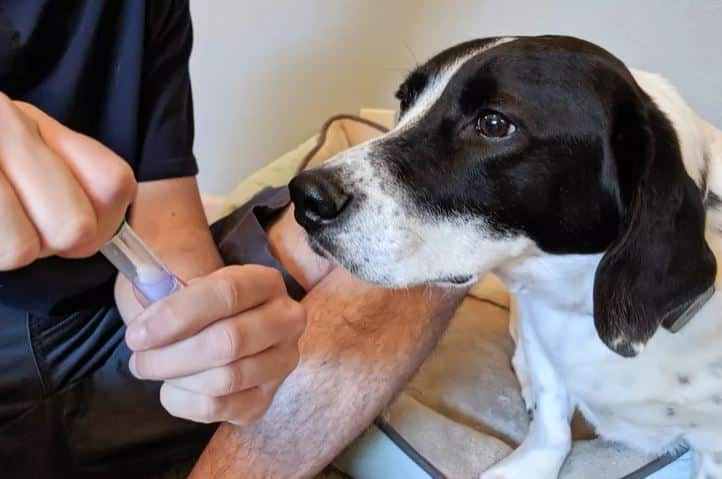
SearchLight DNA™, currently offered through Antech Diagnostics, is a canine cancer genomic diagnostic test that uses predictive biomarkers to provide clinical information about a patient’s tumor to inform treatment decisions. Like a “liquid biopsy” approach, tumor profiling with genomic analysis can provide diagnostic clarity with the benefit of possibly guiding treatment recommendations and prognostic information.
FidoCure™ offers screening of an individual patient’s tumor tissue, identifying specific mutations and translating that into known therapies previously evaluated in dogs with cancer. Confounders of this approach include the identification of mutations of uncertain significance or the fact that there are no existing therapies to target the mutation.
Takeaways
- Utilizing an individual’s cancer genomic signature for selecting specific targeted therapies has proven successful in human oncology, improving outcomes and quality of life for patients with cancer.
- The use of precision medicine in canine oncology is anticipated to grow, leading to better cancer characterization, enhanced therapy selection, and overall improved management of canine cancer.
Artificial Intelligence in Veterinary Medicine
ImpriMed™ offers a novel approach to treating canine lymphoid cancers by developing a personalized treatment protocol for an individual dog with lymphoma. What is referred to as a personalized prediction profile in which each patient is provided with a tailored chemotherapy treatment prediction. This system utilizes artificial intelligence (AI) to generate predictions based on ex vivo drug sensitivity testing and flow cytometry assays.
Components of the ImpriMed Personalized Prediction Profile
Ex Vivo Drug Sensitivity Testing
Tumor cells from the patient are exposed to various chemotherapy drugs in a laboratory setting to determine the sensitivity or resistance of cancer cells to each drug, providing direct evidence of how effective a treatment might be.
Flow Cytometry Assays
This technique identifies specific markers and characteristics of the cancer cells that could influence their response to treatment.
AI Algorithms
The AI algorithms analyze drug sensitivity tests and flow cytometry data to predict the clinical outcomes for different chemotherapy options. This generates a report with personalized treatment recommendations for the oncologist to consider when designing a treatment plan.
The ImpriMedTM prediction profile represents an advancement in veterinary oncology by integrating AI-driven predictions to optimize treatment plans and improve clinical outcomes.
Takeaway
AI-generated reports should always be interpreted in conjunction with the veterinarian’s clinical expertise, patient’s health, and medical history to develop a comprehensive treatment plan.
