-
Adopt
-
Veterinary Care
Services
Client Information
- What to Expect – Angell Boston
- Client Rights and Responsibilities
- Payments / Financial Assistance
- Pharmacy
- Client Policies
- Our Doctors
- Grief Support / Counseling
- Directions and Parking
- Helpful “How-to” Pet Care
Online Payments
Emergency: Boston
Emergency: Waltham
Poison Control Hotline
-
Programs & Resources
- Careers
-
Donate Now
 by Rob Daniel, DVM, DACVIM (Neurology)
www.angell.org/neurology
neurology@angell.org
617-541-5140
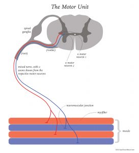
The motor unit is the effector function of those components of the central nervous system responsible for initiating and sustaining movement. This functional ‘unit’ is made up of the lower motor neuron, the nerve rootlet and root, the nerve, neuromuscular junction and the muscle itself.
Dysfunction of the motor unit(s) in a particular limb often shows the hallmark ‘triad’ of clinical signs, namely hyporeflexia, muscle atrophy and hypotonia. The acronym, Neuro-‘RAT’, for Reflexes, Atrophy and Tone has been previously coined (Thomson, Hahn) to help make recognition and recall of such dysfunction easier at the time of a general physical examination.
Once these clinical signs are recognized, the locations of possible disease processes are identical to those of the motor unit, beginning with motor neuron diseases, radiculopathies, peripheral neuropathies, junctionopathies and myopathies.
Once the lower motor neuron system, either in an isolated area or in a generalized area, is identified as dysfunctional, the history, signalment, general physical examination findings and neurologic findings are considered collectively to select appropriate testing, in order to arrive at a diagnosis.
A minimum database (CBC, serum biochemistry and urinalysis) is always indicated in these patients to identify systemic diseases that could be the cause of neuromuscular weakness. Anemia, hypoglycemia, hypokalemia, and hypercalcemia are good examples of diseases that can readily be diagnosed with this testing.
The importance of creatine kinase (CK) testing in these patients cannot be overstated. In consideration of the functional sub-unit of muscle, the sarcomere, CK is an enzyme that acts as a gatekeeper of muscle energy, capable of generating high concentrations of much-needed ATP, when needed. When energy demand is low, this energy is stored as creatine phosphate and when demand increases, this enzyme allows for the rapid restoration of ATP, needed for muscular contraction. This enzyme, a specific marker for myofiber damage, is increased in necrotizing, inflammatory and dystrophic myopathies. Anorexia in cats, along with venipuncture and prolonged recumbency, can also contribute towards elevations of this enzyme activity. Along the same lines, although aspartate aminotransferase (AST), alanine aminotransferase (ALT) and lactate dehydrogenase (LDH) may also be increased with myofiber damage, these enzymes lack the tissue specificity of CK.
Cardiac troponin I, myoglobinuria and lactate/pyruvate testing are often considered in the diagnostic test selection for patients with lower motor neuron disease. For example, cardiac muscle can be involved in a generalized myopathy (e.g. mitochondrial myopathy, necrotizing myopathy) and brown urine (myoglobin in the urine > 250 mcg / mL) often suggests myonecrosis.
Genetic testing has taken off over the past 5-10 years. When faced with generalized weakness in a young purebred dog, possibly involving dysphagia and/or megaesophagus, a quick search of the OFA database and/or Google search can be very helpful, in cases where such a query will suggest a genetic test that can be performed on a DNA sample that can be submitted by mail to the respective lab.
Thyroid function is critical in both energy and lipid metabolism and both hyperthyroidism (in cats) and hypothyroidism should be considered in cases of generalized weakness.
Specific testing, such as acetylcholine receptor antibody testing for Myasthenia Gravis, Type IIM fiber antibody testing in Masticatory Muscle Myositis and muscle/nerve biopsies can be selected at the time of initial testing, considering pattern recognition for these specific diseases. A nerve and/or muscle biopsy can often be instrumental in not only reaching a definitive diagnosis but also in identifying key features of particular disease processes that may have been overlooked at time of initial testing (consideration of mitochondrial and/or lipid storage myopathies).
Electrodiagnostic testing, a supplemental test to the physical and neurologic examination, can often be invaluable, as this is one of the few tests we have at our disposal (like an electroencephalogram, or EEG) that tests function rather than structure (radiographs, computed tomography and magnetic resonance imaging). Lower motor neuron dysfunction, in light of the signalment and history, can often be further explored using this diagnostic modality.
In summary, lower motor neuron disease can often be a challenging but rewarding to treat, depending on where in the disease course the animal presents, the time at which the diagnosis is made and the time at which appropriate therapy is started. Our Neurology service is here at Angell to aid in these challenging cases and may be able to offer further input in regards to diagnostic testing at your hospital.
For more information about Angell’s Neurology service, please call 617-541-5140 or e-mail neurology@angell.org.
References and Useful Links:
Veterinary Neuroanatomy: A Clinical Approach. Christine Thomson and Caroline Hahn. Saunders, Elsevier. 2012
Orthopedic Foundation for Animals: http://www.offa.org/
O’Brien, D.P. and Leeb, T. Invited Review: DNA Testing of Neurologic Disease. J Vet Intern Med 2014; 28: 1186-1198. (Open access)
Shelton, Diane. Invited Review; Routine and Specialized laboratory testing for the diagnosis of neuromuscular diseases in dogs and cats. Veterinary Clinical Pathology. Veterinary Clinical Pathology 39/3 (2010) 278-295.
