-
Adopt
-
Veterinary Care
Services
Client Information
- What to Expect – Angell Boston
- Client Rights and Responsibilities
- Payments / Financial Assistance
- Pharmacy
- Client Policies
- Our Doctors
- Grief Support / Counseling
- Directions and Parking
- Helpful “How-to” Pet Care
Online Payments
Emergency: Boston
Emergency: Waltham
Poison Control Hotline
-
Programs & Resources
- Careers
-
Donate Now
 by Patty Ewing, DVM, MS, DACVP (Anatomic and Clinical Pathology)
www.angell.org/lab
pathology@angell.org
617 541-5014
Definition and Pathophysiology
Bence-Jones proteins, also referred to as M-proteins, myelomas proteins, paraproteins, and free immunoglobulin light chains, are a component of immunoglobulin produced in excess by B cell-derived clonal cell populations. The specific immunoglobulin component is unassociated kappa or lambda light chains. Multiple myeloma and some extramedullary plasma cell tumors and chronic B cell lymphocytic leukemia can produce Bence-Jones proteinuria. Multiple myeloma is a clonal proliferation of malignant plasma cells, typically arising in the bone marrow, that produce an immunoglobulin or component of immunoglobulin. Because of their small size (MW=22,000-44,000), Bence-Jones proteins can readily pass from the blood through the normal glomerular fenestrations of the kidney into the urine. Bence-Jones proteins were named after the English physician Henry Bence Jones, who described their ability to precipitate when urine is heated to 45-70°C, then redissolve when urine is further heated to near boiling.
Considerations for Diagnosis of Multiple Myeloma
Two of the 4 following criteria are generally required for diagnosis of multiple myeloma:
- Radiographic evidence of osteolytic bone lesions
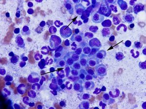
- >20% plasma cells in bone marrow aspirates or biopsy specimens (Figure 1)
- Demonstration of monoclonal or biclonal gammopathy with serum electrophoresis
- Demonstration of Bence-Jones proteinuria
More than 50% of dogs and cats with multiple myeloma may have light chain proteinuria. A diagnosis of multiple myeloma cannot be excluded based on a negative Bence-Jones protein result for the following reasons: 1) secretion may be intermittent or the concentration too low to be detected in a single urine sample, 2) myeloma cells may be secreting intact immunoglobulin molecules rather than free light chains, and 3) non-secretory types of multiple myeloma (rare) do not produce Bence-Jones proteinuria.
Laboratory Tests for Bence-Jones Proteins
Bence-Jones proteins are not detectable via urine protein dipsticks. Detection of Bence-Jones proteinuria requires sophisticated tests such as urine protein electrophoresis, immunoelectrophoresis, and immunofixation electrophoresis. The screening test most commonly used is urine protein electrophoresis.
Sample Requirements for Urine Protein Electrophoresis
Submit 10 ml of urine in a sterile leak proof container or red top (serum) tube on ice packs to the testing laboratory. Twenty-four hour urine collection is ideal, but impractical in most practices. Cystocentesis is the preferred collection method, although catheterized samples and clean free catch are considered acceptable. An early morning urine collection is ideal. Remember to include the method of collection and date/time of collection on the submission form. The urine sample should be kept refrigerated prior to shipping since the proteins are only stable for approximately 2 hours at ambient temperature. The sample is stable at refrigerator temperature (2-8ºC) for approximately one week or can be stored frozen at -18ºC for up to one month.
Interpretation of Test Results
A positive test result will appear as a monoclonal spike in the β or γ protein regions on urine protein electrophoresis. It is important to remember that a negative test result does not exclude a diagnosis of multiple myeloma or B cell neoplasia and a positive result requires additional confirmatory testing. Causes of abnormal test results are presented in Table 1.
Table 1. Causes of Negative and Positive Bence-Jones Test Results
| Positive Result | Negative Result |
|
|
Confirmatory Tests
A positive Bence-Jones protein result on urine electrophoresis can be confirmed and further characterized by more sensitive and specific techniques such as immunoelectrophoresis and immunofixation electrophoresis. Immunofixation electrophoresis distinguishes between kappa and lambda light chains and identifies the heavy chains of IgA, IgM, and IgG. The most common problem reported with immunofixation electrophoresis is that it also detects intact immunoglobulins in the urine that are unassociated with Bence-Jones protein. Additional diagnostic tests that may be helpful in confirming a diagnosis of multiple myeloma include serum protein electrophoresis to evaluate for monoclonal or biclonal gammopathy, radiography to evaluate for osteolytic lesions, and bone marrow cytology to evaluate for plasmacytosis (Figure 1).

Figure 1: Bone marrow cytology from a 10 year old dog with multiple myeloma. Note clusters of plasma cells (black arrows) among the hematopoietic precursors. 500x magnification, Wright-Giemsa stain.
For more information, please contact Angell’s Pathology service at 617-541-5041 or pathology@angell.org.
References
- Bienzle D. Hematopoietic Neoplasia. In: Latimer KS, Mahaffey EA, Prasse KW (eds) Duncan and Prasse’s Veterinary Laboratory Medicine Clinical Pathology, 4th ed., Ames, Iowa: Iowa State Press, 2003: 89-90.
- Thrall MA et al. Laboratory Evaluation of Bone Marrow. In: Thrall MA et al. (ed.), Veterinary Hematology and Clinical Chemistry, Philadelphia: Lippincott Williams & Wilkins, 2004: 172-174.
- Ewing, PJ. Bence-Jones Proteins. In: Vaden SL, Knoll JS, Smith FWK, and Tilley LP (eds) Blackwell’s Five-Minute Veterinary Consult: Laboratory Tests and Diagnostic Procedures: Canine and Feline, Wiley-Blackwell, 2009: 85-87.