-
Adopt
-
Veterinary Care
Services
Client Information
- What to Expect – Angell Boston
- Client Rights and Responsibilities
- Payments / Financial Assistance
- Pharmacy
- Client Policies
- Our Doctors
- Grief Support / Counseling
- Directions and Parking
- Helpful “How-to” Pet Care
Online Payments
Emergency: Boston
Emergency: Waltham
Poison Control Hotline
-
Programs & Resources
- Careers
-
Donate Now
 by Pamela Mouser, DVM, MS, DACVP
Anatomic Pathologist, Angell Animal Medical Center
pathology@angell.org
www.angell.org/lab
617-541-5014
Occasionally, while evaluating biopsy samples, I wonder if certain dog breeds might be better off having their spleens removed at the time of elective sterilization to avoid what seems to be an inevitable future of a ruptured splenic mass, associated hemoperitoneum, and resultant emergency splenectomy ending up as glass slides on my desk. Obviously it is only a fraction of our retrievers and German shepherd dogs that suffer this sequence of events, but it certainly feels more rampant when a rash of spleens is submitted for histopathology.

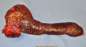
Figure 1: Excised canine spleen with an approximately 8 cm nodular mass. The white boxes illustrate four examples of representative sections which would target the spleen/mass junction.
The decision to perform a splenectomy must be made quickly by some owners, as it is often done on an emergency basis when a splenic mass is actively bleeding. In other cases, owners may have time to consider options when a mass is identified in the spleen incidentally during palpation or ultrasound. In either scenario, a percentage of splenic masses are malignant and are therefore associated with a poor long-term prognosis. The prevalence of malignancy is reportedly higher in cases with hemoperitoneum,1,3,5 but is still significant in non-emergent cases and therefore is a factor in determining treatment options.
Is there a way to know when it is a “bad” splenic mass?
While histopathology is considered the gold standard to determine the nature of splenic mass lesions, distinguishing a benign process from malignancy has been attempted using other diagnostic modalities. I recall as a veterinary student learning an ambiguous generalization that “the larger the splenic mass, the better”, implying that benign processes achieve greater overall size. A study testing this hypothesis found that benign lesions did indeed tend to have a higher mass-to-splenic volume ratio and a higher spleen-to-body-weight percentage than hemangiosarcoma.4 However, the weight measurements did not help in distinguishing benign processes from other types of splenic malignancies (non-hemangiosarcomas). In addition, the measurement could only be performed following removal of the spleen, so it did not preclude surgical intervention. It would be useful if certain clinical or clinicopathologic parameters helped to distinguish the nature of splenic masses. As alluded to above, three retrospective studies found that dogs with non-traumatic hemoperitoneum1,5 and with hemoperitoneum due specifically to splenic masses3 had a greater prevalence of hemangiosarcoma (63.3-70.4%) when compared to dogs in large-scale retrospective studies which included all splenic samples (whether associated with hemoperitoneum or not; prevalence less than 25%).6,7 In the JAVMA study evaluating dogs with splenic masses and hemoperitoneum,3 those patients with hemangiosarcoma also had significantly lower total solids concentrations and platelet counts at the time of admission. Imaging is often employed in the diagnostic workup of these cases. Anecdotally, ultrasonographers are more concerned about the possibility of hemangiosarcoma when a splenic mass appears cavitated; however, I could not find any literature evaluating the predictive value of this finding. In a study focused on computed tomography of splenic masses, the authors noted a significant difference in attenuation characteristics of benign versus malignant processes following contrast administration.2 This technique has not been well-established, and is therefore not routinely applied by radiologists in a clinical setting. Unfortunately, while several factors might increase the clinical suspicion for splenic hemangiosarcoma (signalment, presence of hemoperitoneum, low total solids, cavitated splenic mass on ultrasound, etc), no single feature or combination of findings has been found to adequately predict malignancy. As a result, histopathology is typically necessary.
So then, what are the chances it is a “bad” splenic mass?
In the past 4.5 years at Angell, over 220 spleen samples have been submitted for histopathology. In addition to the typical clinical history of a splenic mass with or without associated hemoperitoneum, less-common submissions include splenic torsions (sometimes associated with gastric dilatation-volvulus) and diffuse splenomegaly. A summary of diagnoses from submitted spleens is included in Table 1. Seven of the spleens were from cats; three of which had mast cell tumors. One spleen was from a ferret and is included in the lymphoma category. All remaining splenic submissions (216) were from dogs. Approximately 46% of canine spleens had a neoplastic process, including a few benign neoplasms. This prevalence is similar to large-scale retrospective studies evaluating splenectomy samples from dogs.6,7 In the Angell population, just over one-third of dog spleens were diagnosed with hemangiosarcoma.
Table 1: Summary of diagnoses for 224 splenic submissions from 2009-2013
| DIAGNOSIS: Neoplasia | |
| Hemangiosarcoma | 75 |
| Other sarcomas | 14 |
| Lymphoma (excludes indolent forms) | 5 (1 ferret) |
| Mast cell tumor | 4 (3 cats) |
| Myelolipoma | 2 |
| Hemangioma | 2 |
| Metastatic anal sac gland carcinoma | 1 |
| TOTAL Neoplasia | 103 |
| DIAGNOSIS: Non-neoplasia | |
| Lymphoid hyperplasia | 51 (1 cat) |
| Hematoma, hemorrhage, +/- extramedullary hematopoiesis | 50 (1 cat) |
| Infarct, torsion, +/- splenitis | 17 (2 cats) |
| Benign fibrohistiocytic nodule | 2 |
| Ectopic bone | 1 |
| TOTAL Non-neoplasia | 121 |
What steps might help optimize the chance of a definitive diagnosis?
A diagnosis of splenic hematoma can cause anxiety for the pathologist, clinician, and owner. I have diagnosed several hematomas in dog spleens without identifying an underlying cause and, while hematomas may occur as a sole lesion, I am always left with the disconcerted feeling that I am missing something important. This is especially the case when other factors are present to increase the suspicion of hemangiosarcoma (i.e. older golden retriever with a cavitated splenic mass and hemoperitoneum). In order to alleviate these fears as much as possible, we follow a few guidelines at Angell in an attempt to capture small or subtle lesions in the midst of a large hematoma. First, representative sections are collected from the junction of the mass lesion and the adjacent unaffected spleen (see Figure 1). Second, areas of different color, texture, and consistency within the mass are sampled. Third, the remainder of the spleen/mass is stored in the pathology morgue in the event that more samples are necessary following initial microscopic evaluation. Some commercial and university laboratories will recommend submitting the entire spleen so that lab technicians or pathologists can evaluate the mass firsthand and select representative sections. Trust me, the pathologist wants to find the underlying lesion causing that giant ruptured splenic hematoma just as much as you do!
TAKE HOME POINTS:
- Neoplastic processes account for almost 50% of all splenic submissions.
- Histopathology is generally necessary for a definitive diagnosis, including distinguishing between benign and malignant processes.
- Representative sections for histopathology should target the junction between the mass and “normal” spleen, and should include areas of differing color, texture, consistency, etc.
For more information, please contact Angell’s Pathology Service at 617-541-5014 or pathology@angell.org.
References
- Aronsohn MG, Dubiel B, Roberts B, Powers BE. Prognosis for acute nontraumatic hemoperitoneum in the dog: a retrospective analysis of 60 cases (2003-2006). J Am Anim Hosp Assoc 2009;45:72-7.
- Fife WD, Samii VF, Drost WT, Mattoon JS, Hoshaw-Woodard S. Comparison between malignant and nonmalignant splenic masses in dogs using contrast-enhancing computed tomography. Vet Radiol Ultrasound 2004;45:289-97.
- Hammond TN, Pesillo-Crosby SA. Prevalence of hemangiosarcoma in anemic dogs with a splenic mass and hemoperitoneum requiring a transfusion: 71 cases (2003-2005). J Am Vet Med Assoc 2008;232:553-8.
- Mallinckrodt MJ, Gottfried SD. Mass-to-splenic volume ratio and splenic weight as a percentage of body weight in dogs with malignant and benign splenic masses: 65 cases (2007-2008). J Am Vet Med Assoc 2011;239:1325-7.
- Pintar J, Breitschwerdt EB, Hardie EM, Spaulding KA. Acute nontraumatic hemoabdomen in the dog: a retrospective analysis of 39 cases (1987-2001). J Am Anim Hosp Assoc 2003;39:518-22.
- Spangler WL, Culbertson MR. Prevalence, type, and importance of splenic diseases in dogs: 1,480 cases (1985-1989). J Am Vet Med Assoc 1992;200:829-34.
- Spangler WL, Kass PH. Pathologic factors affecting postsplenectomy survival in dogs. J Vet Intern Med 1997;11:166-71.