-
Adopt
-
Veterinary Care
Services
Client Information
- What to Expect – Angell Boston
- Client Rights and Responsibilities
- Payments / Financial Assistance
- Pharmacy
- Client Policies
- Our Doctors
- Grief Support / Counseling
- Directions and Parking
- Helpful “How-to” Pet Care
Online Payments
Referrals
- Referral Forms/Contact
- Direct Connect
- Referring Veterinarian Portal
- Clinical Articles
- Partners in Care Newsletter
CE, Internships & Alumni Info
CE Seminar Schedule
Emergency: Boston
Emergency: Waltham
Poison Control Hotline
-
Programs & Resources
- Careers
-
Donate Now
 By Pamela Mouser, DVM, MS, DACVP
www.angell.org/lab
pathology@angell.org
617-541-5014
Lesions occurring in the digit, particularly those associated with the claw and its highly-sensitive supportive structures, can cause substantial discomfort and notable lameness. The digit has unique anatomy in which the bone of the distal or third phalanx (P3) is narrowly separated from the external environment by connective tissue covered by the thick hard keratinized epithelium of the claw. Claws are predisposed to trauma given their natural protrusive orientation. Also, the claw fold “pocket” in which nailbed epithelium gives rise to the keratin lining the claw is an opportune location to entrap foreign material and infectious agents. As a result, both neoplastic and non-neoplastic entities are often clinically significant and produce sufficient morbidity and tissue damage to require amputation of the affected digit. Digit amputation is not only palliative, but can be used for diagnostic purposes at the same time.
In this article, I would like to compare the common diagnoses from digit amputations performed at Angell with those from a previously-published, large multi-institutional study. Our data include 60 amputated digits submitted to Angell Pathology for histopathologic evaluation over the past ~3 years. Of these 60 digits, 50 were canine, 8 were feline, and 2 were from rabbits. A neoplastic process (either malignant or benign) was identified in the majority of the digits (44/60 or 73%), with non-neoplastic entities making up the remaining 16 cases. Table 1 outlines specific diagnoses for amputated digits in dogs from Angell.
| Table 1: Canine digit amputations | |
| Subungual squamous cell carcinoma | 15 |
| Soft tissue sarcoma Osteosarcoma Sarcoma, not further specified Histiocytic sarcoma Total sarcomas |
3 1 2 1 7 |
| Mast cell tumor, grade II | 7 |
| Plasmacytoma | 4 |
| Melanoma | 2 |
| Subungual keratoacanthoma | 2 |
| Cutaneous histiocytoma | 1 |
| Nailbed epithelial inclusion cyst | 4 |
| Chronic pododermatitis, cause not specified Pododermatitis with bacterial paronychia Total pododermatitis |
3 1 4 |
| Onychitis | 2 |
| Fibroadnexal hamartoma | 1 |
| Acral lick dermatitis | 1 |
| TOTAL | 50 |
When our canine data was compared to the large retrospective study1 evaluating canine amputated digits submitted for pathology, subungual squamous cell carcinoma emerged as the most common diagnosis in both study sets (15/50 or 30% of Angell submissions and 109/404 or 27% of digits in the multi-institutional study). At Angell, squamous cell carcinomas were diagnosed in digits from five of six standard poodles, three of three Scottish terriers, and on two separate occasions (two separate digits) from a single giant schnauzer. Other breeds in the Angell group with subungual squamous cell carcinoma included one each of a briard, cocker spaniel, German shepherd dog, Kerry blue terrier, and Labrador retriever. Breeds in which a diagnosis of subungual squamous cell carcinoma was overrepresented in the multi-institutional study included Rottweilers, giant schnauzers, standard poodles, dachshunds, and flat-coated retrievers. Certainly our data supports the overrepresentation of standard poodles and perhaps giant schnauzers with this neoplasm, but we have not recently received amputated digits from the other three breeds reported in that study to make further comparison.
Interestingly, melanoma was the second most common canine digital neoplasm diagnosis in the large-scale study1 (52/404 or 13%) as compared to only 2/50 or 4% of Angell cases. Additionally, the retrospective study found Scottish terriers to be overrepresented with this type of neoplasm, whereas all recent amputated digits from Scottish terriers at Angell (n=3) were diagnosed with subungual squamous cell carcinoma. Both of the digital melanomas diagnosed at Angell occurred in Labrador retrievers.
A greater proportion of inflammatory cases were reported in the published study1 (108/404 or 27%) as compared to digit amputations at Angell (7/50 or 14%). The most common non-neoplastic lesion—excluding inflammation—identified in dog digits from both the retrospective study and at Angell was nailbed epithelial inclusion cyst (20/404 or 5% and 4/50 or 8%, respectively).
Diagnoses from feline digits amputated in the past 3 years from Angell are listed in Table 2. Our number of recent feline digit amputations is likely too low to make many comparisons to the large multi-institutional retrospective study on the same topic.2 However, it is interesting to note that the large retrospective study found squamous cell carcinoma to be the most common neoplastic diagnosis in cat digits, while we do not have a single case of feline digital squamous cell carcinoma on file for the past few years.
Table 2: Feline digit amputations
| Soft tissue sarcoma | 1 |
| Osteosarcoma | 1 |
| Fibrosarcoma | 1 |
| Hemangiosarcoma | 1 |
| Chronic pododermatitis, cause not specified | 1 |
| Pyogranulomatous pododermatitis w/ fungal hyphae | 1 |
| Progressive angiomatosis | 1 |
| Eosinophilic plaque | 1 |
| TOTAL | 8 |
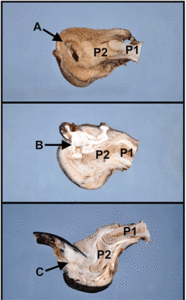
A common theme in cases of digit pathology is the presence of osteolysis affecting the distal phalanx. In the large retrospective study evaluating canine digits1 and based on our clinical experience at Angell, osteolysis can be present in neoplastic, inflammatory, and non-neoplastic/non-inflammatory diseases of the digit. Thus, it is important to remember that radiographic evidence of bone lysis does not necessarily correlate with prognosis. As an example, Figure 1 depicts three amputated digits which have been formalin-fixed, decalcified, and sectioned in a sagittal plane to evaluate the claw-associated lesion as well as phalangeal bones. All three digits have lysis of P3 and include pathology which results in the production of keratin, and therefore can have a similar gross (and perhaps even cytologic) appearance. Histopathology of the amputated digit is often crucial in distinguishing between non-neoplastic, benign, and malignant entities, and is necessary to establish prognosis for a wide variety of conditions.

Figure 1: Three amputated canine digits from Angell, sectioned sagittally to demonstrate each lesion in relationship to the claw (oriented towards the left of each image) and the phalanges. In all images, portions of the first (P1) and second (P2) phalanx are evident, while the distal/third phalanx (P3) is partially to completely effaced by the pathologic processes. The lesions identified by arrows include a nailbed epithelial inclusion cyst (A), a keratoacanthoma (B), and a subungual squamous cell carcinoma (C). In this particular series, the claw is still intact with the squamous cell carcinoma (C) but is blunted to lost in the non-neoplastic and benign processes (A and B).
TAKE HOME POINTS:
- Digit lesions are often painful, especially if osteolysis is present. Digit amputation can be palliative and can be used concurrently for diagnostic/prognostic purposes.
- Osteolysis can occur with a wide variety of digital diseases (neoplastic, inflammatory, and non-neoplastic/non-inflammatory) and therefore does not necessarily correlate with prognosis.
- The most common condition diagnosed in amputated canine digits is subungual squamous cell carcinoma.
References:
- Wobeser BK, Kidney BA, Powers BE, et al. Diagnoses and clinical outcomes associated with surgically amputated canine digits submitted to multiple veterinary diagnostic laboratories. Vet Pathol 2007;44(3):355-361.
- Wobeser BK, Kidney BA, Powers BE, et al. Diagnoses and clinical outcomes associated with surgically amputated feline digits submitted to multiple veterinary diagnostic laboratories. Vet Pathol 2007;44(3):362-365.