-
Adopt
-
Veterinary Care
Services
Client Information
- What to Expect – Angell Boston
- Client Rights and Responsibilities
- Payments / Financial Assistance
- Pharmacy
- Client Policies
- Our Doctors
- Grief Support / Counseling
- Directions and Parking
- Helpful “How-to” Pet Care
Online Payments
Emergency: Boston
Emergency: Waltham
Poison Control Hotline
-
Programs & Resources
- Careers
-
Donate Now
![]()
 x
x
Pamela Mouser, DVM, MS, DACVP
angell.org/lab
pathology@angell.org
617-541-5014
April 2025
x
xx
Bob Barker, the 35-year host of “The Price is Right” and a strong animal rights advocate, would prompt viewers to spay and neuter their pets at the end of each television broadcast. This mantra was subsequently passed to the current host of the game show, Drew Carey. The shift in veterinary medicine to promote pet sterilization, which occurred in the 1970s, is touted as a major factor in reducing the number of animals presented to—and euthanized by—shelters in the decades that followed.1 This widespread practice of sterilizing animals at an early age, which is now commonplace in the United States, likely reduced the number of testes submissions to diagnostic pathologists over time, as gonads were routinely removed prior to developing pathology. While not necessarily the basis of the 1970s sterilization movement, early castration certainly prevents the development of testicular tumors. This article is designed to refresh your memory of testicular tumor types, summarize testicular tumor diagnoses made over a ten-year period at Angell Pathology, and compare Angell tumor types/numbers to published retrospective studies from around the world.
Canine testicular tumors arise from one of two—sometimes both—basic components in the testis: sex cord stromal elements or germ cells.2 Sex cord stromal tumors include Sertoli cell tumors and interstitial (aka Leydig) cell tumors. Seminoma is the predominant germ cell tumor type. In some instances, a single tumor may have both germ cell and sex cord stromal elements, earning the classification of mixed germ cell-sex cord stromal tumor (MGC-SCST). In short, and as a potential spoiler alert for all the upcoming data in this article, dogs with testicular tumors will overwhelmingly have ONE OF THREE TUMOR TYPES: Sertoli cell tumor, interstitial cell tumor, or seminoma. Individual tumors with mixed components (MGC-SCST) or testes containing more than one of these tumor types are not uncommon. Any other tumors, such as the ever-exciting teratoma containing teeth or hair, are remarkably rare.
Canine Testicular Tumors: Pathology
Sertoli cell tumors, which arise from the supporting cells lining the seminiferous tubules, typically appear as nodular to multinodular, white to grey (less commonly tan to yellow), very firm masses. Expansile growth, causing testicular enlargement or distortion, may occur with Sertoli cell tumors (Figure 1C). While malignancy is uncommon, larger tumors may have an increased risk of metastasis. Histologically, neoplastic cells resemble their normal counterparts with elongate columnar to fusiform cell shape and small nuclei, and demonstrate either intratubular or diffuse growth supported by dense fibrous stroma. The fibrous stroma contributes to the firm nature of Sertoli cell tumors grossly, and can be a helpful distinguishing factor for this tumor type. Sertoli cell tumors may get more attention than the other two testicular tumor types as they are the most likely to be hormonally productive, causing a paraneoplastic syndrome of apparent hyperestrogenism. Visually observable clinical signs may include enlarged nipples or gynecomastia, pendulous prepuce, altered behavior around male dogs, contralateral testicular atrophy, and bilaterally symmetric alopecia.2
Interstitial cell tumors, arising from the androgen-producing cells between seminiferous tubules, appear grossly as discrete, yellow, soft nodules with frequent hemorrhage (Figure 1B). These tumors are rarely malignant. Like Sertoli cell tumors, interstitial cells forming masses resemble their normal counterparts with polygonal cell shape, abundant eosinophilic cytoplasm, fine lipid-type vacuolation, and small nuclei. Stroma separating cells into nests and lobules is fine, unlike the dense fibrous stroma seen with Sertoli cell tumors. Hormonal production and paraneoplastic syndromes are considered rare with this tumor type.2
Seminomas are derived from germinal epithelium (spermatogonia) within seminiferous tubules. Grossly, these tumors are homogenous pale tan, grey, or white, bulge from the cut section, and range from soft to moderately firm (Figure 1A, B). The color and consistency bear some resemblance to lymphoma. These masses can become quite expansive, resulting in enlargement or deformation of the affected testis. Similar to Sertoli cell tumors, seminomas can have a tubular or diffuse growth pattern microscopically. Neoplastic germ cells are large, round to polyhedral, and have large nuclei with prominent nucleoli. Variation in nuclear size (anisokaryosis) and multinucleated cells are common, as are mitotic figures, giving all seminomas a malignant histologic appearance despite most tumors having benign behavior.2

Figure 1: Formalin-fixed testes from three dogs. A: Half of each bisected testis with attached epididymis from a 15-year-old golden retriever. The testis to the left is relatively normal to serve as a comparison. The testis on the right is diffusely expanded by a homogenous tan bulging mass, confirmed microscopically to be a seminoma. B: Both bisected testes from an adult Chihuahua. The homogenous tan mass expanding the testis on the left is a seminoma. The discrete nodular yellow mass with focal hemorrhage in the testis on the right is an interstitial cell tumor. C: Half of the bisected left inguinal (cryptorchid) testis from a 5-year-old husky presenting with alopecia and mammary enlargement. The expansile mass distorting the testis is multinodular, tan to yellow with multifocal hemorrhage, and has abundant dense white fibrous stroma. The mass was confirmed microscopically to be a Sertoli cell tumor.
Canine Testicular Tumors: Comparison
A search of biopsies submitted to Angell Pathology from the past ten years resulted in 156 testes from 107 dogs. Of these submissions, 92 testes (59%) had tumors identified; 46 (29%) had non-neoplastic lesions, including processes such as inflammation, vascular compromise, or atrophy; 13 (8%) had no significant microscopic abnormalities; and 5 (3%) did not include identifiable testicular tissue. Dogs with tumors ranged from 4 to 17 years in age. Table 1 lists the number and percentages represented by each tumor type out of the total number of testes evaluated. Note that all testicular tumors evaluated at Angell during the selected timeframe are one of the three types described above, with two mixed tumors containing a combination of germ cells and sex cord stromal elements (MGC-SCST). More than 15% of testes had multiple tumors observed within the same testis. Table 1 also compares Angell’s data with several published retrospective studies from multiple countries worldwide. Note that most studies reported data from a total number of tumors, not the number of testes or dogs, which removes the data point for multiple tumors. Highlighted cells denote the tumor type diagnosed most frequently in each study. The “other” tumors identified in three of the studies included five hemangiosarcomas, four hemangiomas, and one each of granulosa cell tumor, sarcoma, carcinoma, fibrosarcoma, gonadoblastoma, adenomatoid tumor, mesothelioma, lipoma, and metastatic mast cell tumor from scrotal skin.
Table 1: Canine testicular tumors from Angell Pathology compared with published retrospective studies.
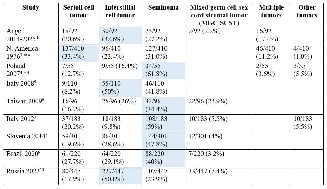
Highlighted cells denote the most common tumor type in each study.
All studies reported data on the total number of TUMORS with the exception of:
*Data reported out of a total number of TESTES.
**Data reported out of a total number of DOGS.
Canine Testicular Tumors: Take-Home Points
- Canine testicular tumors are overwhelmingly one of three types: Sertoli cell tumor, interstitial (Leydig) cell tumor, or seminoma.
- Paraneoplastic signs of apparent hyperestrogenism (feminization, symmetric alopecia, contralateral testicular atrophy) are most often associated with Sertoli cell tumors.
- While Sertoli cell tumors and seminomas may infrequently be malignant, the vast majority of canine testicular tumors are benign and cured by castration.
- While not explicitly discussed in this article, cryptorchidism increases the risk of testicular tumor development.
References
- Rowan A, Kartal T. Dog population & dog sheltering trends in the United States of America. Animals 2018;8:68 org/10.3390/ani8050068.
- Agnew DW, MacLachlan NJ. Tumors of the genital systems. In: In: Meuten DJ ed. Tumors in domestic animals 5th Ames, IA: John Wiley & Sons Inc. 2017:706-14.
- Hayes Jr, HM, Pendergrass TW. Canine testicular tumors: epidemiologic features of 410 dogs. Int J Cancer 1976;18:482-7.
- Sapierzynski R, Malicka E, Bielecki W, et al. Tumors of the urogenital system in dogs and cats. Retrospective review of 138 cases. Pol J Vet Sci 2007;10:97-103.
- Grieco V, Riccardi E, Greppi GF, et al. Canine testicular tumours: a study on 232 dogs. J Comp Pathol 2008;138:86-9.
- Liao AT, Chu PY, Yeh LS, LIN CT, Liu CH. A 12-year retrospective study of canine testicular tumors. J Vet Med Sci 2009;71:919-23.
- D’Angelo AR, Vita S, Marruchella G, Di Francesco G. Canine testicular tumours: a retrospective investigation in Abruzzo and Molise, Italy. Vet Ital 2012;48:335-9.
- Svara T, Gombac M, Pogorevc E, et al. A retrospective study of canine testicular tumours in Slovenia. Slov Vet Res 2014;51:81-8.
- Nascimento HHL, dos Santos A, Prante AL, et al. Testicular tumors in 190 dogs: clinical, macroscopic and histopathological aspects. Pesq Vet Bras 2020;40:525-35.
- Gazin AA, Vatnikov YA, Sturov NV, et al. Canine testicular tumors: an 11-year retrospective study of 358 cases in Moscow region, Russia. Vet World 2022;15:483-7.