-
Adopt
-
Veterinary Care
Services
Client Information
- What to Expect – Angell Boston
- Client Rights and Responsibilities
- Payments / Financial Assistance
- Pharmacy
- Client Policies
- Our Doctors
- Grief Support / Counseling
- Directions and Parking
- Helpful “How-to” Pet Care
Online Payments
Emergency: Boston
Emergency: Waltham
Poison Control Hotline
-
Programs & Resources
- Careers
-
Donate Now
 By Ruth Van Hatten, DVM, DACVR
![]()
angell.org/diagnosticimaging
diagnosticimaging@angell.org
617-541-5139
April 2025
X
X
X
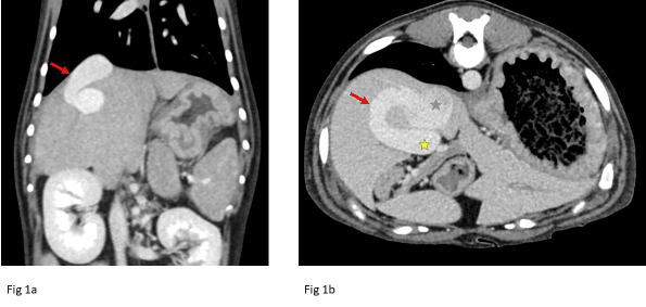
Fig 1a: Example of an intrahepatic portosystemic shunt. Red arrow denotes the shunting vessel. In Fig 1b, the blue star marks the caudal vena cava and the yellow star marks the portal vein.
Computed Tomography (CT) has become a mainstay in veterinary medicine. With the now commonality of this diagnostic tool, it has become vital to make certain distinctions between the various CT protocols. Routine CT scans include a pre-contrast scan followed by administration of contrast medium and a post-contrast scan. This scan is performed to optimize contrast enhancement of the organ parenchyma. Compare this to CT angiography (CTA), which includes a pre-contrast scan followed by post-contrast scans that are timed to achieve specific vascular phases (i.e., arterial, portal, venous, and delayed). CT angiography is the recommended imaging technique to diagnose various vascular abnormalities and characterize various vascular parenchymal lesions.

Fig 2: Example of nest of tortuous vessels (red arrow) secondary to an arterioportal fistula documented in an arterial phase. Due to the secondary portal hypertension, there is a large accumulation of ascites (blue arrow).
One of the most common indications for CTA is the evaluation of portosystemic shunts (PSS). Congenital intrahepatic PSS occurs more frequently in large-breed dogs, while congenital extrahepatic PSS is more common in small-breed dogs. It is recommended to further classify either type of shunting prior to surgical intervention (Fig.1). CTA has been shown to be the best modality for characterizing the origin and termination of the shunting vessel as well as evaluating for possible additional shunting vessels that can be missed on ultrasonography or intraoperative mesenteric portovenography.1 Other portal vascular diseases that can be evaluated on CTA include arterioportal fistulas, acquired portosystemic shunting, and evaluation of portal vein thrombosis, which can mimic portal hypertension secondary to liver disease. CTA is the modality of choice for portal vein disease with improved detection compared to ultrasound (Fig. 2).2

Fig 3: Example of a persistent right aortic arch (red arrow). The green outline denotes a focal, severely distended esophagus with gas and fluid.
Other vascular anomalies that are evaluated with CTA include vascular ring anomalies, coronary artery evaluation, arteriovenous fistulas, and thromboembolism, including pulmonary thromboembolism. Vascular ring anomalies are often present in young patients with regurgitation and focal esophageal enlargement cranial to the heart base. There are various types of vascular ring anomalies, including persistent right aortic arch, aberrant subclavian arteries, and double aortic arch, and they can all present with similar clinical signs. CTA will allow for classification of the type of vascular ring anomaly and surgical planning (Fig. 3). Treatment for pulmonary valve stenosis is balloon valvuloplasty; however, the presence of certain coronary artery anomalies is a contraindication for this treatment and can increase the risk of sudden death during the procedure. CTA of the thorax is required for those breeds with a higher risk of this anomaly to map the coronary artery anatomy prior to the valvuloplasty procedure.3 Evaluation of the arterial phase is required to diagnose an arteriovenous fistula. Thus, a routine CT is not sufficient to diagnose this anomaly. A CTA scan is needed to diagnose thromboemboli, especially in the thorax, where the vasculature cannot be evaluated on ultrasound.

Fig 4: Example of an early post contrast scan. The blue arrow marks a markedly contrast enhancing nodule within the pancreas (red arrow) that was confirmed to be an insulinoma. The black arrows mark metallic hemoclips secondary to a prior liver biopsy.
CTA is also the preferred diagnostic test for evaluating insulinomas. Insulinomas are often small and can be difficult to detect on abdominal ultrasound, especially in large or deep-chested dogs, where the entire pancreas is a baseline difficult to image. It has been documented that insulinomas exhibit increased contrast uptake on the arterial phase on CTA, and then most become similarly contrast enhancing to the pancreas parenchyma on the delayed post contrast phase (Fig. 4).4,5 This imaging characteristic makes it difficult to detect small tumors on routine CT, with CTA being a required diagnostic test for insulinomas. In addition, during the arterial phase, marked contrast-enhancing lesions within the liver and lymph nodes can strongly indicate regional metastasis.5

Fig 5: The red oval marks a large left-sided hepatic mass (presumptive hepatocellular carcinoma). Fig 5a is an early post contrast scan and Fig 5b is the delayed post contrast scan. Note the heterogeneity and large size of the mass in both scans and the more marginal contrast enhancement on 5a.
Lastly, CTA has been used for surgical planning of tumor resection and possibly for evaluating certain tumor types. Using preoperative CTA for isolated hepatic masses has allowed for a more accurate diagnosis of the location within the liver and vascular and multilobar involvement, which can better predict surgical difficulty and resectability. Ultrasonography has been shown to correctly localize solitary hepatic masses in only about 50% of cases.6 The most common malignant hepatic tumor type in dogs is hepatocellular carcinoma, which is often described as a large mass with central cyst-like or necrotic regions on CT, compared to nodular hyperplasia, which is smaller and has a more homogeneous appearance post contrast. On CTA, hepatocellular carcinomas tend to exhibit heterogeneous contrast enhancement with irregular central or marginal contrast enhancement in the arterial phase (Fig. 5). In addition, hepatocellular carcinomas tend to be more hypoattenuating compared to the liver parenchyma on the more delayed phases.7 Nodular hyperplasia, on the other hand, tend to exhibit more homogeneous hyperattenuation or isoattenuation compared to the liver parenchyma and other primary hepatic masses on the various post contrast phases, and hepatic metastatic lesions more commonly are homogeneously hypoattenuating in the earlier post contrast phases.8 Despite these imaging characteristics helping to prioritize malignant vs benign lesions, other reports document more of an overlap between tumor types, and ultimately, histopathology remains the gold standard for diagnosis.9
References
- Parry AT and White RN. Comparison of computed tomographic angiography and intraoperative mesenteric portovenography for extrahepatic portosystemic shunts. 2017;58:49-55.
- von Stade LE, et al. Prevalence of portal vein thrombosis detected by computed tomography angiography in dogs. JSAP. 2021;62:562-569.
- Gunther-Harrington CT, et al. Non-electrocardiographic-gated computed tomographic angiography can be used to diagnose coronary artery anomalies in Bulldogs with pulmonary valve stenosis. 2019;60(1):38-46.
- Coss P, et al. The appearance of canine insulinoma on dual-phase computed tomography angiography. JSAP. 2021;62:540-546.
- Skarbek A, et al. Confirmed and presumed canine insulinomas and their presumed metastases are most conspicuous in the late arterial phase in a triple arterial phase CT protocol. 2023;64(5):834-843.
- Smola C, et al. Computed tomography angiography aids in predicting the resectability of isolated liver tumors in dogs. 2023;261(10). http:/doi.org/10.2460/javma.23.03.0156.pdf Accessed March 13, 2025.
- Fukushima K, et al. CT characteristics of primary hepatic mass lesions in dogs. 2012;53(3):252-257.
- Kutara K, et al. Triple-phase helical computed tomography in dogs with hepatic masses. 2014;55(1):7-15.
- Jones ID, et al. Associations between dual-phase computed tomography features and histopathologic diagnoses in 52 dogs with hepatic or splenic masses. 2016;57(2):144-153.