-
Adopt
-
Veterinary Care
Services
Client Information
- What to Expect – Angell Boston
- Client Rights and Responsibilities
- Payments / Financial Assistance
- Pharmacy
- Client Policies
- Our Doctors
- Grief Support / Counseling
- Directions and Parking
- Helpful “How-to” Pet Care
Online Payments
Emergency: Boston
Emergency: Waltham
Poison Control Hotline
-
Programs & Resources
- Careers
-
Donate Now

 by Megan Whelan, DVM, DACVECC and
Kiko Bracker, DVM, DACVECC
Ultrasound has become an imaging modality that is commonly used in many general practices, specialty hospitals, and emergency rooms. Although considerable training and practice are needed to perform a thorough abdominal ultrasound with accuracy, many emergency rooms are using ultrasound as part of the initial physical examination, as a quick method to evaluate a patient’s abdomen or chest for the presence of free fluid. Little training is required to become quite proficient at this simple evaluation, which can take less than five minutes and can save lives. This article discusses abdominal focused assessment with sonography for trauma (AFAST) and thoracic focused
assessment with sonography for trauma (TFAST).
AFAST was first described in veterinary medicine by Boysen, et al. in 2004.1 It is a technique that we apply to almost every trauma patient in our emergency room, but it also finds use for patients with bleeding abdominal masses, acute abdomen, fluid accumulations from heart failure, and as a daily post-surgical screen in the recovery period following abdominal surgery. In traumatic cases, the AFAST is often repeated after fluid resuscitation to look for free fluid accumulation, and to see if the urinary bladder is filling with urine. Animals having an anaphylactic reaction can show gall bladder wall thickening, with striations that can be detected on an AFAST exam.2
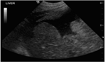 |
| Figure 1: A large volume of neoplastic effusion in a cat, likely due to a pancreatic mass. Liver lobes are visible. |
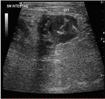 |
| Figure 2: Small volume of a cellular effusion (ingesta) next to the small intestine in a dog. The dog had been bitten by another dog, rupturing the bowel. |
To perform the AFAST exam, the patient is positioned in either right or left lateral recumbency and then four specific sites are scanned in a fanned fashion with the probe (Figure 3). The first site is the diaphragmatico-hepatic location (1), which evaluates the area of the liver, stomach, and diaphragm; second is the spleno-renal location (2), which evaluates the spleen and the right kidney; third is cysto-colic region (3), which evaluates the area of the urinary bladder; and lastly is the hepato-renal location (4), which focuses on the right kidney and the right side of the liver.1,3 The patient’s positioning, the four specific locations of evaluation, and the order they are evaluated are described differently by different sources. Using the same procedure with each patient is helpful in order to compare patient to patient, and to become familiar with the location and structures within the abdomen. When bleeding is severe, the most dependent location is often where free abdominal fluid is located and can then be sampled via abdominocentesis. When bleeding is milder the hemorrhage is found near the location of the damaged organ.3
 |
| Figure 3: Used with permission by Gregory R. Lisciandro, DVM, Dipl. ABVP, DACVECC |
The abdominal fluid score (AFS) is used to rate the severity of blood/fluid accumulation using the AFAST technique and it follows a simple four-point scale. If fluid is present at one of the four sites, a point is given. An AFS of 0 means there is no abdominal fluid, whereas an AFS of 4 means there is fluid present in all four sites using the AFAST technique.3 Dr. Gregory R. Lisciandro determined that patients with an AFS of 1–2 rarely become anemic, whereas patients who have an AFS of 3–4 often become anemic and about 25% of them require blood transfusions.4
The FAST technique can also be applied to the thorax. TFAST permits evaluation for pneumothorax, ”wet” or “dry” lungs, or more simply for pleural or pericardial fluid.5 Patients can either be in lateral or sternal recumbency, but both sides of the thorax should be evaluated. Pleural fluid often accumulates just caudal to the heart (and rostral to the diaphragm). This is also a convenient place to perform thoracocentesis because the depth of the fluid is often greatest at this location, making damage to the lung, heart, or other intrathoracic structures during thoracocentesis unlikely. When pericardial fluid is present, the heart/pericardium is usually very easy to image because it is larger and often comes closer to the chest wall, making interference from the air-filled lung less likely.
 |
| Figure 4: Pericardial effusion in a cat. The nearly anechoic effusion is visible between the pericardium (visible as a white arcing line at the bottom of the view) and the bright epicardium that is seen on the surface of the myocardium. |
While the physical exam is still where the most important information is gleaned about a patient, the FAST techniques can be used in many situations to give additional information and further clarify the clinical picture of a traumatized or critically ill patient.
For more information about Angell Animal Medical Center’s Emergency/Critical Care service, please visit www.angell.org/emergency. To contact Angell’s E/CC doctors by phone or to refer a patient to the Angell E/CC service, please call 617 522-5011 or e-mail emergency@angell.org. You can also reach Dr. Bracker at kbracker@angell.org and Dr. Whelan at mwhelan@angell.org.
- Boysen SR, Rozanski EA, Tidwell AS, et al. Evaluation of a focused assessment with sonography for trauma protocol to detect free abdominal fluid in dogs involved in motor vehicle accidents. J Am Vet Med Assoc 2004; 225(8):1198–1204.
- Quantz JE, Miles MS, Reed AL, White GA. Elevation of alanine transaminase and gallbladder wall abnormalities as biomarkers of anaphylaxis in canine hypersensitivity patients. J Vet Emerg Crit Care 2009; 19(6):536–544.
- Lisciandro GR, Lagutchik MS, Mann KA, et al. Evaluation of an abdominal fluid scoring system determined using abdominal focused assessment with sonography for trauma in 101 dogs with motor vehicle trauma. J Vet Emerg Crit Care 2009; 19(5):426–437.
- Lisciandro GR. Case-Based Applications of Abdominal (AFAST) and Thoracic (TFAST) FAST techniques for Trauma, Triage, and Tracking (Monitoring). Syllabus from New England Regional Veterinary Conference, Sept 23–25, 2011, Portland, ME.
- Lisciandro GR, Lagutchik MS, Mann KA, et al. Evaluation of a thoracic focused assessment with sonography for trauma (TFAST) protocol to detect pneumothorax and concurrent thoracic injury in 145 traumatized dogs. J Vet Emerg Crit Care 2008; 18(3):258–269.