-
Adopt
-
Veterinary Care
Services
Client Information
- What to Expect – Angell Boston
- Client Rights and Responsibilities
- Payments / Financial Assistance
- Pharmacy
- Client Policies
- Our Doctors
- Grief Support / Counseling
- Directions and Parking
- Helpful “How-to” Pet Care
Online Payments
Emergency: Boston
Emergency: Waltham
Poison Control Hotline
-
Programs & Resources
- Careers
-
Donate Now
 By Terri Bright, Ph.D., BCBA-D, CAAB
![]()
angell.org/behavior
behavior@angell.org
617-989-1520
January 2022
Dogs Are . . . Animals
Our dogs are our family members, yet we rarely think of them as animals, saying to ourselves, “I have an animal living in my house!” Thus we are often surprised when left to their own devices, dogs behave…like animals. They eat gross things. They chew our best shoes. They eat homework. If you were to ask them, “Why?” they might paraphrase Sir Edmund Hillary: “Because they were there!” Luckily, it is not too hard to keep them occupied and trained by following some simple rules, using special equipment, and learning a little more about your best friend’s world.
From Pups to Adults – What to Use and How to Train
Young puppies need to be exposed to the world at a young age, even before they have had all of their vaccinations; in fact, this is the recommendation of the American Veterinary Society of Animal Behaviorists [1]. Called “socialization,” the weeks of 4-14 are a specific time in a dog’s life when they can most easily learn to tolerate noises, scary things, different types of people, and how to behave appropriately with other dogs. Ideally, if a puppy looks frightened of something, they should be allowed to move away from it while being fed some treats. They should not go to the wild and crazy dog parks because they may earn to fight if frightened, but instead should be paired with other dogs who will be gentle with them and who have similar play styles.
Whether you have a puppy or an adult dog, training is essential. But yikes, how do you know where to start? It’s actually easy: find out what they love, and pay them with that when they do what you want them to. Here is a hint: dogs have a tremendous sense of smell, so anything that smells terrible will probably act as a good reward. Freeze-dried meats are terrific, especially because one treat can be the size of half of a pea.
There are some standard behaviors to train that can be helpful: the obvious are sit, down, stay, come when called, and loose-leash walking. Simply give the dog a treat immediately after they do what you ask. You may have to use the treat to get them started for a new behavior, but make sure to quickly change over to giving the treat afterward, so you do not have to bribe them into doing what you ask. Stay on the side of the angels when training, always rewarding when the dog gets it right, and never frightening them by shaking something loud, using collar corrections or electronic collars. The side effects of scaring your dog are well known in behavioral literature [2] and include fear and aggression. If you need training help, join a rewards-based group class such as we have in Boston, Waltham, and Methuen (www.mspca.org/dogtraining). Remember that even if your dog is well-behaved in your kitchen, when the environment is more exciting, e.g., anywhere outside the kitchen, much practice will be required to make the behavior fluent everywhere.
Helpful Equipment
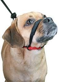
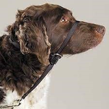
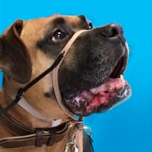
Helpful equipment for strong dogs who have not learned to walk on a loose leash includes head harnesses such as the Sporn, Gentle Leader, or Snoot Loop (pictured below, left to right).
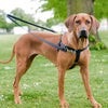
Our favorite body harness is called the Freedom No-Pull Harness (pictured below), which comes with a unique leash so you can hook the leash to the front and top rings. If you “pay” your dog a treat at your left side and feed them every two to four steps, they will hang out at your left side more often.
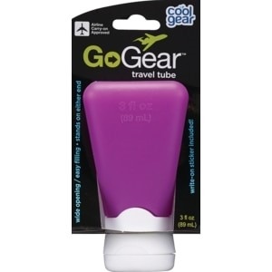
A great addition to your training bag is a 2- to 3-ounce silicone squeeze bottle (pictured below) sold at drug stores for lotion or shampoo. It is easy to fill it with peanut butter or light cream cheese, even wet dog food, for a long-lasting treat you can deliver with gloves on.
Enrichment
As it turns out, a leashed march around the neighborhood in heel position is not your dog’s idea of a good time. They have 200 million odor receptors (we have a measly 5 million), so they are actually a nose on four legs. On a walk, they smell who/what peed where, the track of a squirrel or cat, even who walked by carrying a pizza. It’s fun to find a balance of their walking with you…and you’re following them on their crooked scent trail. Other doggish things your dog will thank you for: toss a handful of their kibble in the grass for them to sniff out. (In the winter, you can hide kibble around the house for them.) A “snuffle” mat is another place to hide treats; it looks like a shag rug. A “licki” mat is a rubber dish you can smear peanut butter on for your dog to lick while you trim their nails or brush them.
Other favorite food-delivery toys are the Omega Tricky Treat Ball and the Snoop- no need to put kibble in a bowl! You can set your dog’s wet food in a Kong and freeze it for a long-lasting meal for your dog.
Your canine family member is devoted to you. You can pay them back – one treat at a time – to change their behavior and feed them in a way that is doggedly inspiring to them. What better way to enhance your wonderful and mysterious bond with them?
References
- https://avsab.ftlbcdn.net/wp-content/uploads/2019/01/Puppy-Socialization-Position-Statement-FINAL.pdf
- https://avsab.ftlbcdn.net/wp-content/uploads/2021/08/AVSAB-Humane-Dog-Training-Position-Statement-2021.pdf