-
Adopt
-
Veterinary Care
Services
Client Information
- What to Expect – Angell Boston
- Client Rights and Responsibilities
- Payments / Financial Assistance
- Pharmacy
- Client Policies
- Our Doctors
- Grief Support / Counseling
- Directions and Parking
- Helpful “How-to” Pet Care
Online Payments
Emergency: Boston
Emergency: Waltham
Poison Control Hotline
-
Programs & Resources
- Careers
-
Donate Now
 By Courtney Peck, DVM, DACVECC
![]()
angell.org/emergency
MSPCA-Angell West
781-902-8400
Intravenous fluid therapy has been a cornerstone of medical and surgical management in human, as well as veterinary medicine for decades. The primary goal behind using intravenous fluids is to maintain or restore intravascular volume, tissue perfusion, and reverse dehydration. Different types of fluids (such as crystalloids, natural or synthetic colloids, or blood products) have different compositions in terms of tonicity, acid-base, oncotic properties, hemostatic and immunologic effects. These differences translate into varying effects in our patients.
 Historically, fluids were thought to operate within the parameters established by Starling’s forces; where hydrostatic pressure worked to push fluids out of the vasculature, and oncotic pressure worked to retain fluid within. More recently, this theory has been updated after the identification of the endothelial glycocalyx, a meshwork of long glycosaminoglycans (GAGs) linked to other proteins (glycoproteins and proteoglycans) that lines the luminal surface of the vasculature. Identification of this structure has led to a revision of Starling’s forces, as we have realized the significant role of the glycocalyx in how the body responds to fluid therapy (and explains why our fluid plans don’t always do what we think they are going to do). The hydrostatic and oncotic pressure gradients between the microvascular lumen and the interstitium rely heavily on the integrity and health of the endothelial glycocalyx.
As advances in understanding fluid therapy and the physiology behind its use occur, many debates and disagreements have developed in both human and veterinary medicine about fluid therapy. Standardized fluid therapy recipes (a.k.a. “one size fits all”) are no longer supported; instead, current recommendations urge that fluid therapy is detailed for the individual patient, taking into consideration their clinical illness, needs/deficits, and comorbidities. Recommendations for continued evaluation of our patients while on fluid therapy, and using goal-directed resuscitation, are currently in favor.
So how do we incorporate this into our daily practice?
Fluid Distribution in the Body
Before considering how to incorporate fluid therapy, we need to back up and refresh ourselves on how fluids are distributed in the body. Approximately 60% of our body is water; this is referred to as our total body water (TBW). Water plays many roles in the body, including transport of electrolytes, nutrients, and blood cells; carrying substrates across membranes; evaporative cooling; and participation in metabolic functions and reactions. In general, we think about two main fluid compartments within the body: the intracellular fluid (ICF) and the extracellular fluid (ECF). The intracellular space consists of 40% of our TBW, and the intracellular space comprises roughly the other 15 to 30%. The extracellular space is further divided into the interstitial component (which is 75% of the ECF) and the intravascular component (which is 25% of the ECF). Most of the intravascular fluid is plasma. You can remember this breakdown by using the 60:40:20 rule – TBW is 60%, divided as 40% ICF and 20% ECF. In order to create a tailored fluid plan for a patient, we need to understand and remember how fluid is distributed in these different compartments.
The different body fluid compartments are separated by membranes. The endothelial membrane and endothelial glycocalyx separate intravascular and interstitial space, while cell membranes separate the ICF from the ECF. Fluid movement across the different compartments depends on membrane characteristics, hydrostatic pressure, colloid oncotic pressure, and osmolarity.
Fluid Balance and Imbalance
In a normal healthy state, fluid balance is maintained between the various compartments as described above. Normal physiology also normally maintains fluid balance between our patients and their environment. Healthy animals mostly obtain fluids through drinking and eating, as well as a small gain of water via metabolic reactions. They lose water through panting as well as urination and defecation. In certain disease states, such as fever, gastrointestinal disease (decreased intake of water/food, vomiting, diarrhea), respiratory disease, and hemorrhage, additional fluid losses occur.
When disruptions in fluid balance occurs, disorders in hydration and perfusion develop. Most commonly, fluid loss will exceed fluid intake; however in some cases, such as with congestive heart failure, there can be a positive fluid balance. Evaluating a patient’s fluid balance incorporates physical exam findings, diagnostic results, and assumptions based on patient history and signalment. Determining from which compartment(s) a patient has lost fluid is an important first step in determining what type of fluid therapy they may need.
Hydration Versus Perfusion
It is helpful to remember this distinction when considering fluid therapy in our patients. Hydration status refers to TBW and the role of this fluid in tissue support and cellular processes. We assess this through skin turgor, mucus membrane moisture or lack thereof, and ocular position. Dehydration occurs through decreased water intake relative to fluid losses. Fluid loss with dehydration results in interstitial fluid loss as well as intracellular fluid loss. Interstitial fluid loss occurs through standard dehydration, as well as changes in the interstitial colloid oncotic pressure (fluid moves from the intravascular space into the interstitium), resulting in increased plasma sodium and hemoconcentration. Intracellular fluid loss can occur as well.
Perfusion refers to the intravascular volume, which plays a role in providing oxygen and other nutrients to the tissues. Hypoperfusion results from loss of fluid from the intravascular space; most commonly this occurs with acute hemorrhage, however severe dehydration will eventually result in hypoperfusion as well. The body can normally shift fluid volumes to temporarily compensate for small or controlled losses from the intravascular compartment, but if the losses are significant, clinical signs of shock will develop. These include tachycardia, hypotension, delayed CRT and depressed mentation.
A patient who is dehydrated has different fluid therapy needs from a patient who is experiencing hypoperfusion. Once you have identified which type of fluid loss you are dealing with, you can start to make a fluid therapy plan. Fluid therapy also has different phases, depending on what you goal is. Fluid resuscitation refers to correcting perfusion abnormalities, while rehydration refers to correcting hydration abnormalities. Fluid maintenance refers to ongoing fluid therapy once dehydration and/or hypoperfusion have been corrected. Evaluating your goals for a patient will help further determine your fluid therapy plan. Is the patient in shock? Dehydrated? Both? Can your patient drink and eat? Enough to maintain any losses? Is your goal with fluid therapy to correct clinical dehydration or achieve endpoints of resuscitation (normal heart rate, improved blood pressure, CRT < 2 seconds; mentally appropriate)?
Why should we care about fluid therapy?
While there are many obvious benefits of fluid therapy, ongoing research in human and veterinary medicine have suggested that fluid therapy (and specific types of fluids) may have unwanted or adverse effects. By tailoring a fluid plan to an individual patient’s needs, we reduce the risk of adverse effects and increase positive outcomes. Having established goals prior to starting fluid therapy will help guide your decisions and provide objective data on whether you are achieving those goals or need to come up with a different plan. Goals can include correcting dehydration, correcting electrolyte abnormalities, improving blood pressure. In human medicine, targeted end points of resuscitation have improved patient outcome.
Fluid Basics and Choices
In general, there are 4 main categories of fluids: crystalloids, colloids, blood products, and hemoglobin based oxygen carriers (oxyglobin). Transfusion medicine is beyond the scope of this lecture, and HBOCs have been unavailable in the US for years, so I will not cover them either. However they may make a comeback at some point so it is prudent to remember they may someday be potentially useful. As such, in clinical practice, we tend to have crystalloids and colloids to choose from when creating a fluid plan.
Crystalloids
Crystalloids remain the mainstay of fluid therapy in human and veterinary medicine. These fluids contain small solutes (usually electrolytes, <500g/mole), which allows them to easily cross the capillary endothelium into the ECF. Crystalloids are categorized into balanced or unbalanced solutions. Balanced solutions have an electrolyte composition similar to the ECF, while unbalanced (such as 0.9% NaCl) don’t. Crystalloids can also be categorized based on whether they are replacement or maintenance solutions. Replacement solutions (Normosol-R, Lactated Ringers Solution, Plasmalyte-148) are also called isotonic crystalloids, are ideal for correcting fluid deficits in a patient. Maintenance solutions (Normosol-M, Plasmalyte-56) tend to have lower sodium and higher potassium concentrations than the ECF, and are best for providing fluid therapy to replace fluid lost through normal physiologic functions. (I would say in general, while there are a variety of types of crystalloids, we tend to use replacement fluids only, because as soon as a patient is able to replace their normal physiologic fluid losses with eating and drinking, they are off of fluid support).
Crystalloids are attractive because they are readily available, relatively inexpensive, have a long shelf life, are easy to use, and we are all pretty comfortable using them. However, there are a few things to keep in mind clinically, and this all goes back to what your goal with fluid resuscitation is.
When it comes to using crystalloids in fluid resuscitation, it is important to remember that crystalloids have the largest effect on the interstitial and intracellular compartments, due to their rapid equilibrium throughout the ECF. After 30 minutes, half of the volume you deliver remains in the vasculature. This means that while there may be a beneficial immediate effect of blousing crystalloids, their effect is going to be lost rapidly, which may require repeated dosing, which may result in fluid overload of your patient. In general, large volumes of crystalloids are needed to achieve goals of resuscitation. Adverse effects with using such large volumes include dilutional coagulopathy, damage to the endothelial glycocalyx, hypothermia, and fluid overload.
Silverstein et al compared the effects of multiple types of fluids on blood volume, and the results are reflected in this chart (see slides). They used 0.9% saline as the crystalloid representative, and you can see that there is a very nice increase in blood volume in the first 10 to 15 minutes, but that by 30 minutes, the effect is back down to about a 20% increase. Crystalloids provide a rapid volume expansion, but then have a rapid redistribution out of the vasculature.
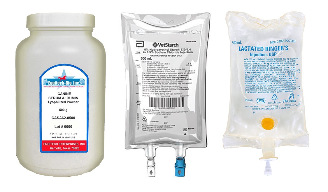
Colloids
When faced with the short-lived effects and potential adverse effects of large volume crystalloid use, creation of colloids became popular. Colloids are fluids which contain large molecular weight solutes (> 10k Da), and can be categorized as natural (such as albumin, some blood products) or synthetic (such as Hetastarch or Vetstarch which contain synthetic starch compounds). This type of fluid tends to remain within the intravascular space due to these large molecules, which prevent it from crossing the (healthy) endothelium. As they remain in the vasculature, they exert a “pulling” effect on fluid in the interstitium, drawing it into the vasculature and keeping it there, thus increasing intravascular volume. This effect allows significant volume expansion without having to deliver a large volume of colloid fluid, because they utilize fluid that is already inside the patient. This volume expansion is also longer lasting that that of crystalloids. In theory, you can volume resuscitate with a smaller amount of colloids than you can with crystalloids. However, these theories have not been supported by human data.
Synthetic colloids are hydroxyethlstarches that are categorized based on their molecular weight, their concentration in solution, and the degree of hydroxyl group substitutions at positions C2 and C6 of the molecular structure. These differences are relevant in light of more recent concerns about adverse effects of synthetic colloid use; some types of synthetic starches are more commonly associated with complications. Those synthetic colloids with longer half-lives, higher molecular weights, and a higher degree of C2/C6 substitution have been associated with higher complication rates. Synthetic colloids are eliminated by the reticuloendothelial system as well as renal elimination. Currently it is believed that these systems will reach a saturation point, and then any remaining synthetic starches in the body create problems.
Synthetic colloids were heavily used when they first became available in human and subsequently veterinary medicine, and they seemed to solve the problems experienced with crystalloid use. However over time, concerns about the safety of colloid use in human (followed by veterinary) medicine came to light, creating some controversy and confusion. Human studies implicated induced coagulopathy, acute kidney injury, and increased mortality following use of synthetic colloids. Veterinary medicine has also looked at any relationship between synthetic colloids and these adverse effects, and the results have not been as compelling.
Coagulation effects: Morris (2016) found prolongation of clotting times in vitro with both LRS and HES, however the HES effects were more severe. Helmbolt (2014) evaluated the effect of HES 670/075 on platelet closure times in vivo (dogs), and found that various CRI rates (1 mL/kg/hr and 2 mL/kg/hr) significantly increased PCT, however there was no evidence of spontaneous bleeding in any of the dogs. Wurlod (2015) performed an in vitro study comparing the effects of multiple crystalloid and synthetic colloid fluids on coagulation, and found that while synthetic starches affected coagulation parameters, they were no more coagulopathic at clinically relevant doses as compared to saline.
Renal effects: In humans, hydroxylethylstarches (HES) were labelled with a black box in 2013 in the US, and banned in the EU, due to concerns over their association with development of acute kidney injury in humans. Since then, synthetic colloid use has largely fallen out of favor; with human albumin becoming a popular alternative. On the veterinary side, Hayes (2016) evaluated the incidence of AKI and death following HES administration in dogs over a 3 year period, and found HES use was associated with an increase in mortality and AKI. These results caused many in the veterinary E/CC realm to rethink our previously liberal use of synthetic colloids. Yozova (2016) also evaluated the association of HES use with creatinine and mortality in dogs, and found that dogs receiving HES had a higher mortality, but no difference in creatinine or the development of AKI. They concluded that outcome was not associated with HES use. Boyd (2019) evaluated changes in renal histopathology after the use of gelatin (another synthetic starch) and HES and found that a urinary biomarker of inflammation (uMCP) was increased after gelatin and HES use compared to crystalloids. Zersten (2019) also evaluated HES use in anesthetized dogs and did not find evidence that HES increased creatinine (in fact creatinine decreased with HES administration). The jury is still out in veterinary medicine as to how much of a clinical risk synthetic colloids pose in our patients; in general their use has dropped off and some loose clinical guidelines have surfaced, including avoiding the use of HES in any azotemic patient or patients with other risk factors for AKI, and limiting the amount of HES to 20 mL/kg/day.
Albumin
Albumin is the most abundant natural colloid; it provides more than 70% of colloid oncotic pressure. Albumin also plays roles in wound healing, coagulation, transportation of hormones, drugs, cations (such as calcium), free radical scavenging, and also acts as a weak buffer. It is the most common colloid used in human medicine, which has allowed a pretty easy move away from synthetic colloids. Human albumin has a 79% homology with canine albumin, and the use of human albumin in veterinary medicine has been reported albeit with some significant adverse effects. Hypersensitivity reactions have been reported in multiple veterinary studies, and the risks have made veterinarians wary of using this product routinely. Canine albumin is a tempting alternative, because theoretically the risk of hypersensitivity reactions would be far less. However thus far, availability and cost have prevented propelling the use of canine albumin into routine use. Lyophilized canine albumin is sporadically available, which makes it an appealing alternative due to a prolonged shelf life, but at this point it is just not consistently available. Fresh frozen plasma and frozen plasma also contain albumin, however the volume of these products needed to significantly increase colloid oncotic pressure is usually cost prohibitive and poses a risk for fluid overload (70 mL/kg/day).
Blood Products
Discussing all of the various blood products is beyond the scope of this lecture, but newer research and protocols in human medicine are pretty interesting and may be clinically applicable to our patients. Dividing whole blood into its components has been a long standing practice in human and veterinary medicine. Over the past 10 years, recognition of phenomena such as dilutional coagulopathy and acute traumatic coagulopathy are forcing us to rethink our resuscitation strategies. The most current transfusion recommendation is to transfuse one unit of fresh frozen plasma and one unit of platelet concentrate for every one unit of packed red blood cells. It seems that compartmentalized transfusion medicine is not as easy or simple as we had hoped, which has caused the pendulum to swing back towards reconsidering the benefits of whole blood transfusions. On the battle fields, they now have people who serve as whole blood donors instead of everyone being transfused components. So while not every veterinary practice may have the space or finances to be able to stock blood products, having that big friendly young dog that is otherwise healthy (and screened) may be an effective way to be able to provide a whole blood transfusion.
Monitoring While on Fluid Therapy
As mentioned above, fluids therapy should be tailored for each individual patient based on their previous medical history/comorbidities, current illness, and other ongoing needs. Likewise, our patients’ physiology and condition change from day to day (or sometimes more frequently), so it is important that we reevaluate them and adjust our fluid therapy plan frequently. It’s great if fluid therapy works out as planned; however it’s just as likely that the initial plan will need to be revised at some point.
When evaluating a patient in light of fluid therapy, physical examination can be very useful. Do they appear euhydrated? Do they appear interstitially overhydrated (jiggly SQ) but hypovolemic (hypothermic, dumpy, hypotensive)? Are they still dehydrated? Using objective parameters such as changes in body weight, degree of urine production, blood pressure, heart rate, as well as laboratory data such as PCV/TS, electrolytes (sodium in particular) and renal values are the best way to assess changes in hydration and perfusion status. Based on these evaluations, you can then adjust your fluid rate or type, and add any supplementation (such as potassium) as indicated. Prolonged fluid therapy when a patient has improved and doesn’t require fluid support can be detrimental, so remembering to taper fluids is also important.
Case Examples
The Dehydrated but Euvolemic Patient
These are the patients that haven’t been eating or drinking normally, or have increased losses to some degree. These patients may be quiet but are responsive, normothermic, and their vital parameters are usually within normal limits. The goal with these patients is to replace their ECF. Since they do not have decreased perfusion at this point, an isotonic, replacement crystalloid is a great choice, because the goal is to get that fluid out of the vasculature and into the interstitium. Start by calculating their daily fluid requirement; there are a few ways to do this. Current recommendations for maintenance fluid rates for dogs are 60 mL/kg/day, and for cats are 50 mL/kg/day. An alternate calculation (which I tend to use for larger dogs because the above calculation often will overestimate their requirements) is based on RER: 30(weight in kg) + 70 = daily fluid requirement. If a patient is markedly dehydrated, you can start with 1.5x the maintenance rate and then evaluate in 6 to 8 hours.
The Shocky, Hypovolemic (not hemorrhaging) Patient
These patients usually suffer from a combination of decreased ECF and intravascular volume, and their physical exam parameters reflect this. These patients are often mentally dull, markedly dehydrated (prolonged skin tent, sunken eyes), bradycardic (cats especially) or tachycardic, hypothemic, and hypotensive. The goal with these patients is to restore intravascular volume quickly, but also replace their ECF fluid loss as well. These patients need fluid replacement more urgently than the above example, so a fluid bolus is usually warranted (caution with preexisting cardiac disease or a murmur/gallop). When thinking about fluid boluses, remember to use the largest IV catheter possible, use more than one if you need to give a large volume (i.e. large dogs), and ideally place the catheter(s) as close to the heart as possible (cephalic veins or even external jugular veins in some cases). The amount of the bolus depends on the patient status. In general, we start by calculating the patient’s blood volume. Historically we referred to this as their “shock volume,” however this term has fallen out of favor as we have reevaluated the impact of fluid therapy in our patients (especially the more critical ones). In dogs, this is 90 mL/kg. In cats, it is 60 mL/kg. It is currently recommended to start with ¼ of the patient’s blood volume, and then reassess. This volume can be repeated if needed, but continual reassessment is imperative. By starting with smaller volumes, we reduce the risk of fluid overload. Once your patient’s clinical parameters (heart rate, body temperature, pulse quality/blood pressure, mentation) improve, you can transition to an initial replacement fluid rate as described above.
The Shocky, Hypovolemic, Hemorrhaging Patient
These patients can present similarly to the above cases, but they usually were euvolemic and euhydrated prior to the hemorrhage event. Their main problem is acute blood loss and secondary shock. We don’t necessarily want our fluid therapy to be lost to the ECF in about 30 minutes; we want it to stick around to support the cardiovascular system and provide perfusion to organs. In these cases, starting with hypertonic crystalloids (such as hypertonic saline) makes sense. Hypertonic saline (HTS) has a high osmolarity (2400 mOsm/L, compared to LRS which has an osmolarity of 273 mOsm/L), which creates an osmotic gradient into the vasculature. Because HTS is a crystalloid, it will also leave the intravascular space rapidly (in about 30 minutes), so HTS should be followed by another type of fluid. Colloids historically have been used in this role, as they will tend to keep all that intravascular volume you pulled from the interstitium in the vasculature longer. HTS should be avoided in profoundly dehydrated patients, or patients who can’t regulate their total body water. It can be used in mild to moderate dehydration, but needs to be followed with isotonic crystalloids to allow fluid to move back into the interstitium. HTS (3-4 mL/kg) should be given intravenously over 15 minutes, due to a risk of vagal response to rapid volume expansion.
As discussed earlier, colloid fluid therapy was extremely popular in these types of cases due to its oncotic support, but use of colloids has decreased over the past 5 years due to safety concerns. Colloids are still helpful for “low volume resuscitation,” where they are combined with HTS (3-4 mL/kg HTS with 4 mL/kg colloid), and can be quite effective in head trauma cases.
Another option in these types of cases would also be blood components (again in a 1:1:1 ratio ideally), or whole blood.
Human and animal studies have shown that survival decreases with large volume crystalloid resuscitation in uncontrolled hemorrhage, so this should be avoided. Large volumes of crystalloids can dislodge clots and dilute coagulation factors. Instead, using “controlled resuscitation” or “hypotensive resuscitation” is recommended in acute trauma patients. This entails giving fluids to achieve a MAP of 40 mmHg (normal MAP ~ 80 mmHg) on a short term basis, until hemorrhage can be definitively stopped. Human studies evaluating this strategy revealed decreased coagulopathies, decreased blood loss, and improved splanchnic perfusion. Another popular strategy in human medicine is “delayed resuscitation,” where fluid resuscitation is withheld until definitive control of hemorrhage is achieved (a.k.a. surgery). The applicability of this strategy to veterinary medicine remains unknown, as we tend to not rush every trauma case into surgery immediately.
The Oliguric/Anuric Patient (Most Commonly Renal Disease Patients)
This category of patients highlights the importance of reassessing your fluid therapy strategy over time. Most animals in renal failure (whether CKD or AKI or acute exacerbation of CKD) are dehydrated, and require fluid therapy to reestablish acid-base and electrolyte derangements, as well as restore hydration. Historically, renal failure cases needed “fluid diuresis,” to remove the uremic toxins and restore renal function. These patients would be automatically placed on up to twice maintenance fluid rates for days. Many of these patients became fluid overloaded and did not survive. When renal function is compromised, urine production is altered (sometimes polyuria, more commonly oliguria or anuria). It is extremely important to monitor the urine output in these patients, because if they are unable to produce urine to the degree that we expect them to, they are at high risk for fluid overload. Additionally, increasing intravascular volume tends to cause some degree of interstitial edema, and this occurs in the kidneys as well. When the kidneys become edematous, renal hypertension can also develop, resulting in a further decrease of urine output. In these cases, it is recommended to create a fluid therapy plan to rehydrate them, not overhydrate them, and monitor their urine output closely. Place a urinary catheter if needed to truly measure urine output, and adjust your fluid therapy accordingly once rehydration is complete. In patients that are euhydrated and remain oliguric with increasing azotemia, or develop anuria, more aggressive fluid therapy will only increase the risk of fluid overload. In these cases, intervention with hemodialysis should be considered as this is the only modality that can stabilize the anuric or oliguric azotemic patient.
Conclusions/Take Home Points
- Fluid therapy is not a cook book, one size fits all strategy; individual patient needs, losses, and parameters should be considered and the fluid therapy plan should be created accordingly.
- Continual reassessment and adjustment of your fluid therapy plan is essential.
- More crystalloids do not improve outcome, and can sometimes make things worse.
- Caution with colloids! Especially in coagulopathic or azotemic patients.
- Whole blood is coming back into favor.
- Pay close attention to urine output in patients with renal failure.